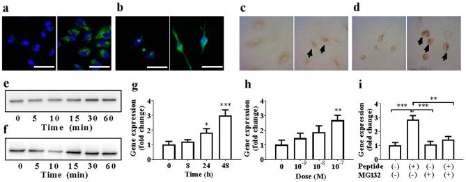
Figure 2.
Cellular responses to GIP_HUMAN[22–51]. (a, b) Confocal laser-scanning microscopy images of the fluorescent GIP_HUMAN[22–51] peptides bound to cultured cells. Growing HAoECs (a) or THP1-derived macrophages deprived of serum for 16 h (b) were overlaid without (left panels) or with 10–6 M FAM-GIP_HUMAN[22–51] (right panels) for 30 or 5 min, respectively. Cells were washed, fixed, nuclei counterstained with DAPI (blue) and the cell surface-bound green fluorescence visualised. Scale bar represents 50 μm. (c, d) Nuclear translocation of NF-κB. HAoECs (c) or THP1-derived macrophages (d) were incubated without (left panel) or with 10–6 M GIP_HUMAN[22–51] for 60 min, and immunocytochemical staining was performed using an NF-κB p65 subunit antibody to detect its nuclear translocation. (e, f) Degradation of IκB-α. HAoECs (e) or THP1-derived macrophages (f) were stimulated with 10–7 M GIP_HUMAN[22–51] for the indicated times and subjected to western blot analysis using the anti-IκB-α antibody to assess the time-course of IκB-α degradation. The panels show the cropped blots and the full-length blots are presented in the Supplementary Information. (g–i) Upregulation of MMP8 gene expression by GIP_HUMAN[22–51]. HAoECs were incubated with 10–7 M GIP_HUMAN[22–51] for the indicated times (g) or with indicated doses for 48 h (h), and MMP8 mRNA and β-actin levels were quantified. Data represent the fold changes (mean ± S.E.M) of MMP8 mRNA copies relative to β-actin mRNA (n = 5). *p < 0.05, **p < 0.01, ***p < 0.001 compared with vehicle. (i) HAoECs pre-treated with or without MG132 (500 nM) for 30 min were incubated with 10–7 M GIP_HUMAN[22–51] and MG132 (100 nM) for 24 h and MMP8 and β-actin mRNA levels were quantified. **p < 0.01, ***p < 0.001, compared with vehicle. The relative mRNA levels are shown as fold changes (mean ± S.E.M; n = 5).

