Figure 6.
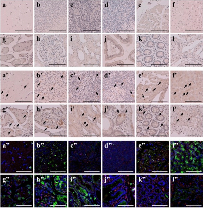
Systemic expression of GIP_HUMAN[22–51] and its negative colocalisation with GIP in major human organs. (a–l, a’–l’) Human tissue array sections were immunohistochemically stained with either IgG purified from preimmune rabbit serum (a–l) or anti-GIP_HUMAN[22–51] IgG at 1:500 dilution (a’–l’). Arrows indicate the positions of positive immunoreactivity. (a”–l”) Confocal microscopic images of immunofluorescent dual staining of GIP_HUMAN[22–51] and GIP. Human tissue array sections were double-stained with either the rabbit polyclonal anti-GIP_HUMAN[22–51] antibody (× 1000) or the mouse monoclonal anti-human GIP antibody (× 500). The red signals corresponding to the localisation of GIP were obtained with the Alexa Fluor 594 secondary antibody (× 1000) and the green signals representing GIP_HUMAN[22–51] were obtained with the Alexa Fluor 488 secondary antibody (× 500). The nuclei were counterstained with DAPI (blue). Overlay resulted in yellow signals indicative of colocalisation. (a–a”) cerebral cortex, (b–b”) cerebellum, (c–c”) tonsil, (d–d”) lymph node, (e–e”) heart, (f–f”) liver, (g–g”) stomach, (h–h”) kidney (glomerulus), (i–i”) kidney (tubule), (j– j”) small intestine, (k–k”) colon and (l–l”) testis (scale bar: 100 μm).

