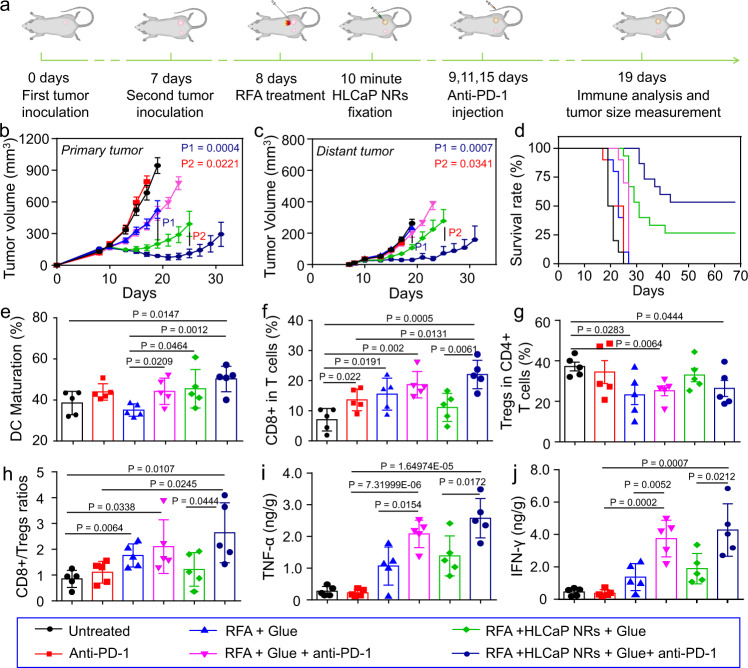
Fig. 6. In vivo antitumor study and corresponding immune mechanism study of combined RFA, HLCaP NRs fixation, and anti-PD-1 immunotherapy.
a Schematic illustration of the inoculation of the bilateral tumor model for in vivo antitumor and immune mechanism studies. b–d Tumor growth curves of primary (b) and distant tumors (c), as well as corresponding mobility-free survival rate (d) of mice with bilateral tumor models post different treatments as indicated. The mouse was set as dead when its tumor volume was larger than 1000 mm3. e DC maturation status in the drain lymph nodes adjacent to the primary tumors post various treatments as indicated. f–h The frequencies of CD3+CD8+ T cells (f), and CD3+CD4+FoxP3+ Tregs (g), as well as their ratios (h) inside the distant tumors post various treatments as indicated. i, j The secretion levels of TNF-α and IFN-γ inside the distant tumors post various treatments as indicated. Data in Fig. b, c were represented as mean ± SEM, n = 10 or 15 biologically independent animals, data in Fig. e–j were represented as mean ± SD, n = 5 biologically independent animals. P values calculated by the two-tailed student’s t-test are indicated in the figure.

