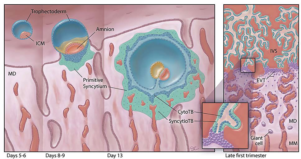
Fig. 1:
First trimester human placental development (left to right). The human blastocyst (far left), comprises an inner cell mass (ICM) completely surrounded by a layer of trophectoderm (TE) cells, and resides within the endometrial cavity just prior to implantation [days 5–6 post-fertilization (pf)]. At the time of implantation (days 8–9 pf), the ICM has developed the beginnings of an amniotic cavity (amnion) and the leading edge of the implanting embryo is characterized by an inner group of proliferative cytotrophoblast cells (CytoTB) and a deeper, non-proliferative, invasive, multinucleated, thickened mass of primitive syncytium. By day 13 pf, the embryo is fully implanted in the maternal decidua (MD). The primitive syncytium now completely surrounds the embryo. Primary villi have begun to form as invaginations of the cytotrophoblast layer. The primitive syncytium can invade the uterine glands (UG) and contacts maternal vessels. It contains lacunae that will grow and coalesce; they are filled with endometrial secretions and small amounts of maternal blood. By late in the first trimester (right panel), the villous placenta has been established with maternal blood now filling the intervillous space (IVS). Villi contain a discontinuous layer of proliferative CytoTB surrounded by multinucleated villous syncytiotrophoblast (SyncytioTB). The anchoring villi extend across the IVS to attach to the MD at their tips. Extending from these anchoring villi are the HLA-G-positive extravillous cytotrophoblast cells (EVTB). EVTB will invade deeply into the MD and remodel maternal spiral arteries. Although not depicted here, they will also invade into maternal veins, uterine glands and lymphatic spaces and reach well into the uterine myometrium. Multinucleated trophoblast giant cells (Giant cell) can be found deep in the MD and in the maternal uterine myometrium (MM).

