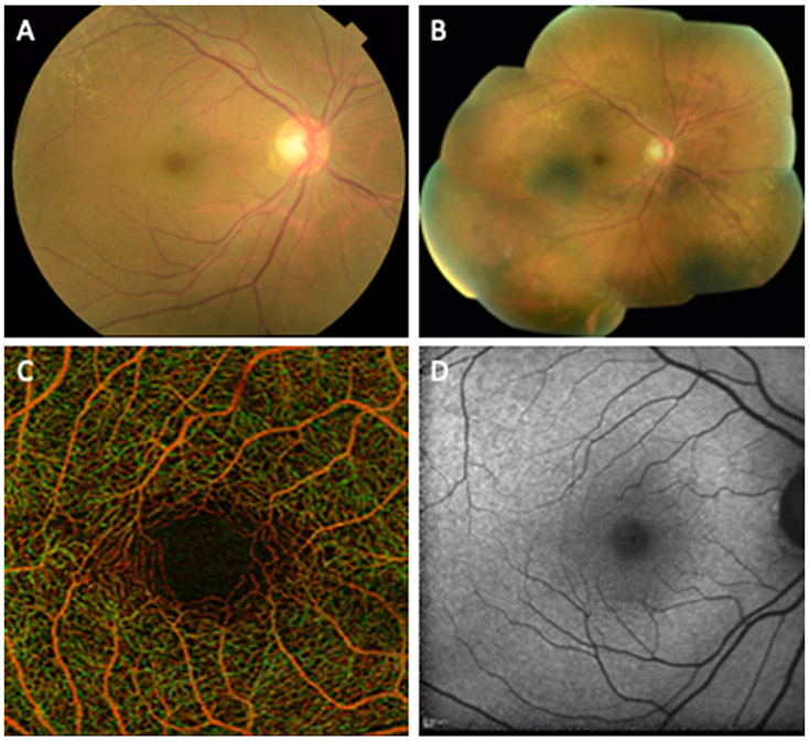
Fig. 1.
Examples of retinal imaging modalities from a 65 year old female illustrate commonly used methods for evaluation of retinal disease and retinal changes in neurodegenerative diseases. (A) Color fundus photograph illustrating the macula, optic disc and retinal arteries and veins. (B) Digital collage of color fundus images of the same subject demonstrating 60-90° field of view that includes the peripheral retina outside the vascular arcades. (C) Optical coherence tomography angiogram of the parafoveal area illustrating the capillaries in the area and the foveal avascular zone. Red and green pseudocoloring represent the depth of retinal capillaries in the superficial and deep retinal layers, respectively. (D) Short wave fundus autofluorescence image of the macula.

