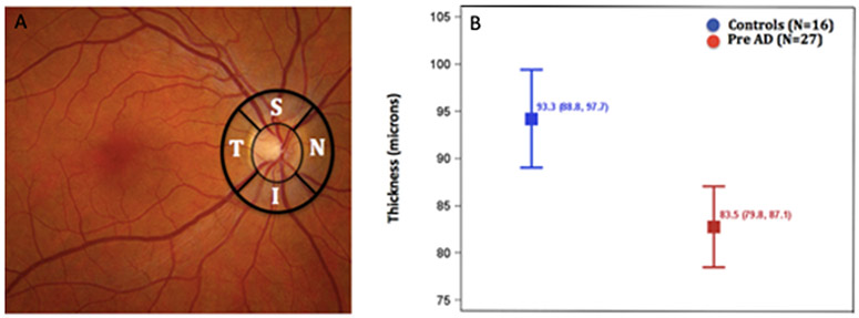
Fig. 5.
Peripapillary retinal nerve fiber layer thickness is reduced in subjects with preclinical AD. (A) The thicknesses of the retinal nerve fiber layer (temporal, superior, nasal, inferior) was measured using OCT in the regions outlined in black. (B) Depicts least-squares mean (95% CI) total retinal nerve fiber layer thickness adjusted for side and region between cognitively healthy controls (blue) and cognitively healthy participants with pathologic CSF Aβ42/Tau levels (red). (Adapted from Asanad S, Fantini M, Sultan W, Nassisi M, Felix CM, Wu J et al. (2020) Retinal nerve fiber layer thickness predicts CSF amyloid/tau before cognitive decline. PLoS ONE 15(5): e0232785. https://doi.org/10.1371/journal.pone.0232785 under the terms of the Creative Commons Attribution License http://creativecommons.org/licenses/by/4.0/).

