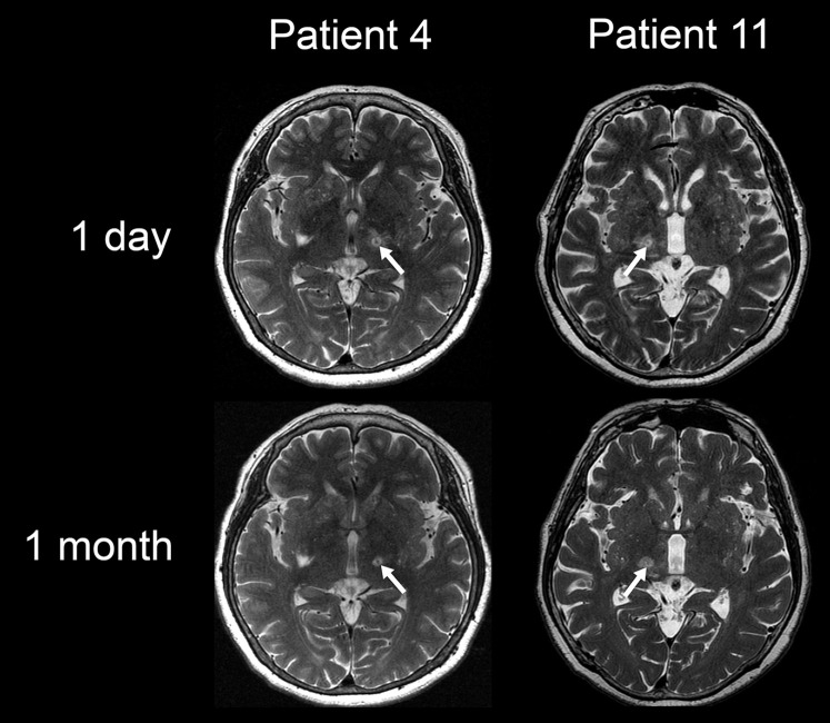
Fig. 2.
T2-weighted magnetic resonance imaging of demonstrative cases. In the case with dysesthesia remaining at 1 year (Patient 11), the lesion showed lateral extension, which was more evident at 1 month than the postoperative day. In contrast, the demonstrative case without any adverse events (Patient 4) showed a lesion staying in a round shape at 1 month. (White arrows represent lesions created by focused ultrasound thalamotomy.)

