Fig. 2.
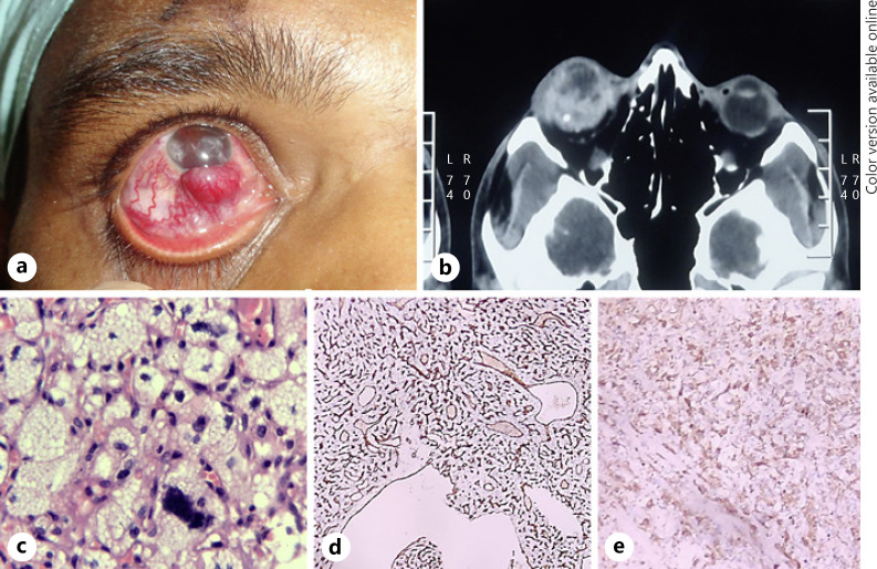
Clinical, imaging, and histopathological findings of case 2. a Vascularized right subconjunctival mass at the inferior limbus with dilated episcleral blood vessels. b CT scan orbits showing ill-defined heterogeneous intraocular mass with areas of calcification within. c Histopathology of the enucleated eyeball (H and E staining, ×10 magnification) showing intraocular mass, abundant vascularity with both thick-walled and thin-walled large vessels, and an extensive capillary network and stromal cells are arranged in lobular patterns with abundant foamy cytoplasm and irregular, hyperchromatic nuclei (H and E, ×40 magnification). IHC staining with CD34 highlighted the capillary network of tumour vasculature (d) and inhibin was positive in stromal cells (e). IHC, Immunohistochemistry; CT, computed tomography.

