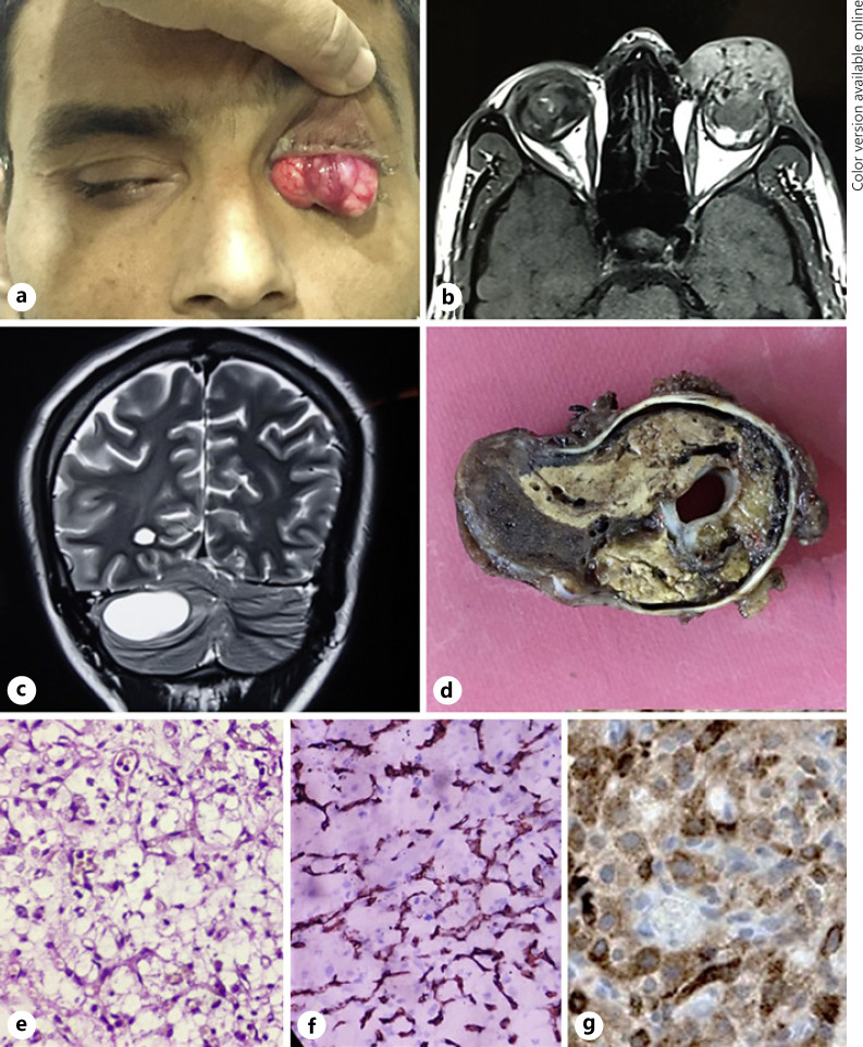
Fig. 3.
a Clinical, imaging, and histopathological findings of case 3. Left eye fungating intraocular mass with extraocular extension. b T1W MRI orbits showing left eye intraocular mass with extraocular extension with flow voids within. c MRI brain with contrast showing cystic mass in left cerebellum suggestive of cerebellar hemangioblastoma. d Gross examination of the enucleated eyeball showing a tan-brown intraocular mass with extraocular extension anteriorly. Histopathology (×40 mag, H and E staining) showing an infiltrative tumour with an anastomotic network of delicate capillary-like blood vessels and conspicuous multivacuolated to foamy appearing stromal cells (e) and CD 34 positivity in vascular walls (f) and inhibin positivity in foamy cells (g).

