
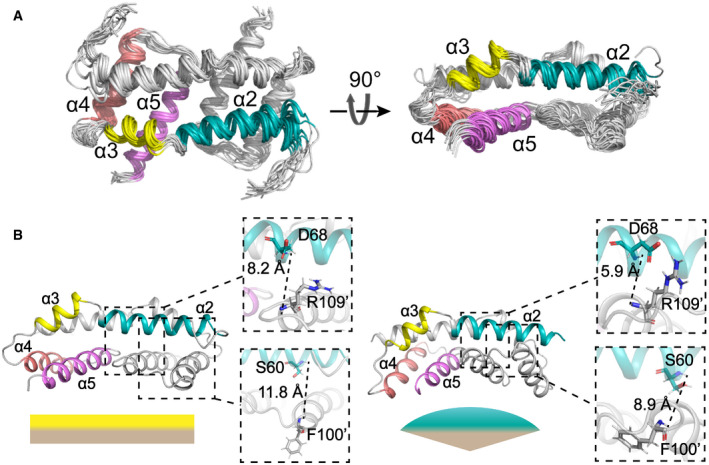
Figure 2. Structures of Bax (α2–α5) in lipid bicelles determined by NMR.
-
AEnsemble of 15 lowest energy structures for Bax (α2–α5) dimer in bicelles calculated from the geometry constraints obtained from the NMR experiments summarized in Table 1. The backbone structures are shown as thin ribbons.
-
BComparison of the NMR structure of Bax (α2–α5) in bicelles (left; PDB code: 6L8V) and the crystal structure (right; PDB code: 4BDU). The NMR or crystal structure has a flat or concave bottom surface formed by the α4–α5 regions, respectively, as indicated by the complimentary flat or convex shapes underneath. The zoom‐in regions display the distances between the amide protons of D68‐R109’ and S60‐F100’, which are longer in the NMR structure than the crystal structure.