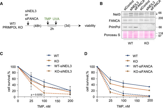
Figure EV6. PRIMPOL and NEIL3 are not epistatic for ICL repair.

-
AExperimental design of CellTiter‐Glo viability assays in WT and PRIMPOL KO cells 3 days after treatment with different doses of TMP for 2 h followed by irradiation with UVA (90 s).
-
BImmunoblots showing the levels of Neil3, FANCA, and PrimPol proteins in WT and KO cells after siRNA‐mediated downregulation of NEIL3 or FANCA. The arrow indicates the band corresponding to Neil3 protein. Ponceau S is shown as loading control.
-
C, DCell survival assays with WT and PRIMPOL KO cells following NEIL3 (C) or FANCA (D) downregulation with siRNA after a 2 h exposure to increasing amounts of TMP (followed by UVA irradiation, 90 s). The control curves with WT and PRIMPOL KO cells without Neil3 or FANCA downregulation are the same as in Fig 7G. Each point represents the percentage of surviving cells (average and SD of three [NEIL3] or two [FANCA] assays). Statistical analysis was conducted with two‐way ANOVA followed by Bonferroni post‐test. The P‐value of PRIMPOL KO vs KO‐siNEIL3 comparison is indicated.
Source data are available online for this figure.
