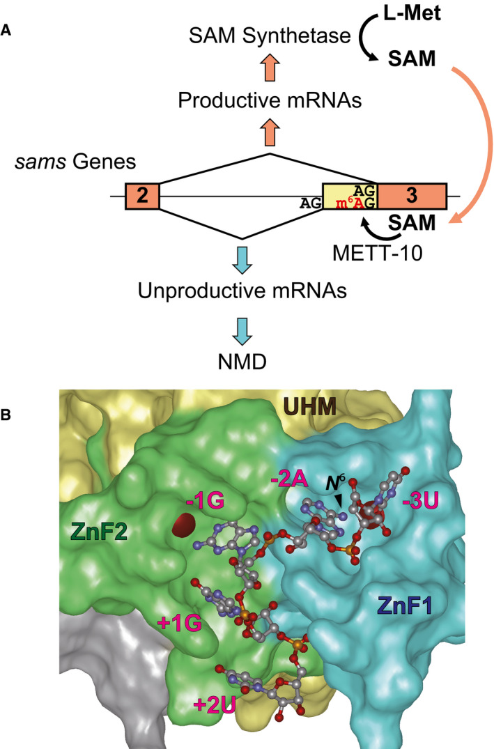
Modeled structure of
C. elegans UAF‐2 binding to 5′‐UAGGU‐3′. The structure was modeled based on sequence homology to U2AF23 from
S. pombe (Appendix␣Fig
S9) and crystal structure of
S. pombe U2AF23/U2AF59 complex bound to the RNA (Yoshida
et␣al,
2020). N‐terminal Zn finger 1 (ZnF1), U2AF homology motif (UHM), and C‐terminal Zn finger 2 (ZnF2) domains are colored in blue, yellow and green, respectively. Red spheres indicate zinc ions. The position of the amino group methylated upon m
6A modification (
N
6) is indicated with an arrowhead.

