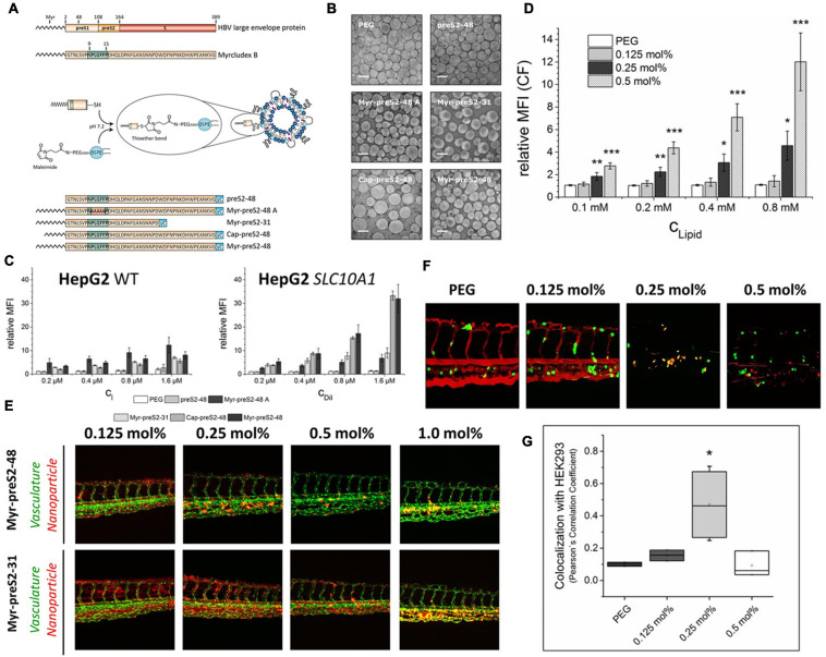
FIGURE 2.
Identifying different peptide ligands for a better NTCP-specific targeting. (A) Schematic representation of HBV large envelope protein, Myrcludex B and its five derivatives. Peptides were conjugated to the end of PEG chain integrated in the liposomes through the connection of sulfhydryl group with maleimide group. (B) The morphological characters of different Myrcludex B-derived lipopeptide conjugated liposomes. (C) The uptake rate of different peptide conjugated liposomes into HepG2 wild type cells and SLC10A1 overexpressing HepG2 cells was identified by flow cytometry analysis. (D) The uptake of liposomes modified with different amounts of Myr-preS2-31 identified by flow cytometry analysis. Significance (*p < 0.05, **p < 0.01, and ***p < 0.001) was calculated relative to PEG, respectively. (E) The performance of Myr-preS2-48- and Myr-preS2-31-modified liposomes was tested in zebrafish embryos expressing green fluorescent protein in their vasculature endothelial cells (green signal). DiI (red signal) was used to label the membrane of liposomes. (F) Myr-preS2-31-modified liposomes were tested in wild type zebrafish embryos xenotransplanted with human, GFP expressing HEK293 cells (green signal), expressing SLC10A1. DiI (red signal) was used to label the membrane of liposomes. Yellow signals demonstrate the colocalization of liposomes with HEK293-GFP cells. (G) Pearson’s Correlation Coefficient (PCC) for quantitative analysis of liposomes binding to HEK293-GFP cells (Witzigmann et al., 2019). Significance (*p < 0.05) was calculated relative to PEG. Open Access.

