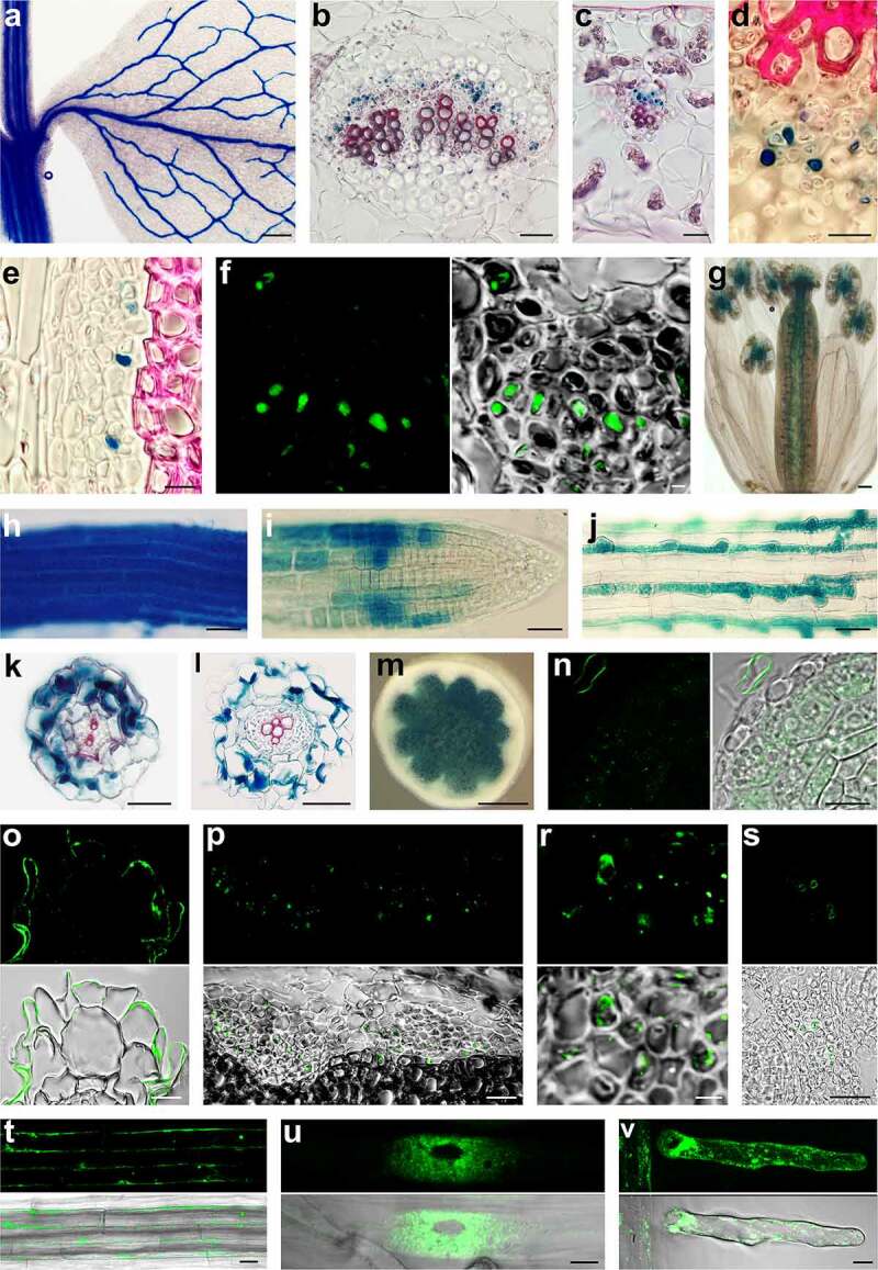
Figure 1.

Gus histochemistry and IRT1 protein localization. (a): Young shoot segment with developing leaf of pIRT1::GUS line. The GUS histochemistry reaction resulted in blue color is visible in the vasculature of leaf and stem. (b) Cross-section through leaf major vein and (c) minor vein of the pITR1::GUS plant shows that IRT1 promoter is active in the phloem part of the vasculature. (d) Detail view of phloem in pITR1::GUS plant leaf vein and stem vascular bundle (e) showing GUS activity in the companion cells and feeble reaction in sieve elements. (f) Immunohistochemical localization of GUS protein in phloem companion cells of pITR1::GUS plant using the antibody against GUS. (g) IRT1 promotor is active in pistil and junctions between filaments and anthers. Maturation (h) and meristem (i) zone of the root of pIRT1::GUS seedlings growing without iron. (j) Signal at the beginning of mature root zone of pIRT1::GUS seedlings when growing on iron supplemented medium is mainly detected in trichoblasts. Cryo-sections through maturation (k) and elongation (l) zone of pIRT1::GUS roots demonstrate the activity of IRT1 promotor in epidermis and cortex but not in the central cylinder. (m) Hand section of the stem of pITR2::GUS line showing GUS activity in the pith. (n) Immunolocalisation of IRT1 protein on cryosections in meristematic zone of roots of wild seedlings growing on iron-free medium. Labeling is seen in intracellular patches but not at the cell periphery. Note labeling at the periphery of root hair of another root accidentally appearing in the vicinity during embedding. (o) IRT1 labeling with IRT1 antibody on cryosection through the maturation root zone detect signal mainly at the periphery of root epidermal cells. (p) Histoimmunology assay on cryosections shows cells labeled with IRT1 antibody scattered in the phloem part of the vascular bundle, and detailed view (r) shows the signal in the phloem companion cells. (s) In pistil, IRT1 was located at the periphery of several cells in the septum transmitting tract. Parts of ovules are on the left and right sides. (t) Long version of IRT protein with attached Dendra2 at its C-terminus is localized at the periphery of epidermal cells in matured root zone and in intracytoplasmic bodies. (u) Short IRT version of IRT1 with Dendra2 attached to its C-terminus is localized to numerous patches surrounding the nucleus (u, image prepared with Image J program represent Z projection of 3 slices) and patches at the periphery as seen in root hair (v, image represent Z projection of 5 slices)
Scale bars: 500 µm (a and m); 100 µm (g); 50 µm (b,c,h,i,j,k,l and p); 25 µm (d,e,n,o,s,t and u); 10 µm (f and r).
