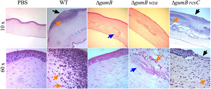
FIG 6.
Histological analysis of S. marcescens infected corneas at 48 h. Representative images of hematoxylin- and eosin-stained corneal sections of eyes at 48 h postinfection. Large neutrophil infiltrates were observed in the WT- and ΔgumB rcsC mutant-infected eyes. The objective magnification is indicated. Black arrows indicate epithelial defects, blue arrows indicate globular infiltrates in ΔgumB mutant-infected corneas, and orange arrows indicate neutrophils.

