Figure 3.
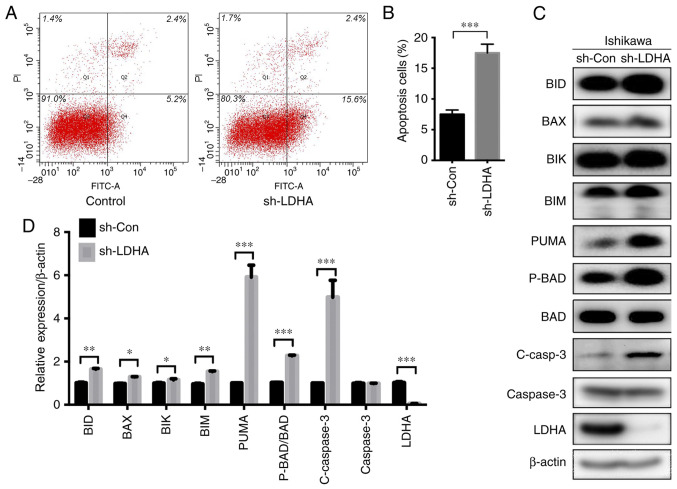
Effect of LDHA on apoptosis of Ishikawa cells. (A) Representative graphs of apoptosis detected by flow cytometry. (B) A higher number of Ishikawa cells underwent apoptosis when LDHA was knocked down (sh-LDHA) compared with the control (sh-Con). (C) Western blot analysis of apoptosis-related proteins. (D) Histogram of BID, BAX, BIK, BIM, PUMA, P-BAD/BAD, C-caspase 3, total caspase 3 and LDHA protein expression in Ishikawa cells. Values are expressed as mean ± SD of three independent experiments. Student's t-test was used to compare two groups. *P<0.05, **P<0.01 and ***P<0.001. LDHA, lactate dehydrogenase A; C-, cleaved; sh-, short hairpin; Con-, control; P-, phosphorylated.

