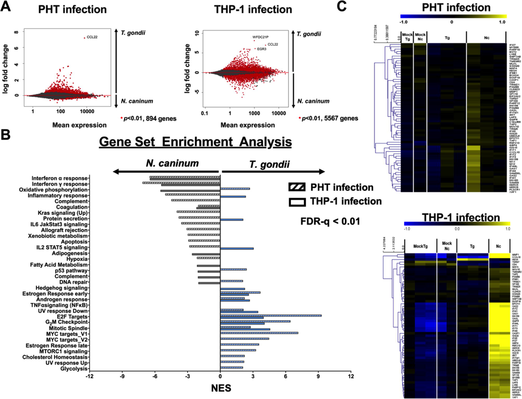
Fig. 1.

Differential expression analysis of human trophoblast cells (PHTs) against Toxoplasma gondii and Neospora caninum tachyzoite infection. (A) Plots showing mean expression against log fold change of the transcriptomic profile of PHTs during T. gondii and N. caninum infections. Each dot represents a host gene and genes that are significantly different in response to the parasitic infection (in comparison to mock infection) are represented by ● (P<0.01). (B) Gene set enrichment analysis of the PHT (solid bar, either blue or gray) and THP-1 (hatched bar, either blue or gray) transcriptomes in response to T. gondii (blue) and N. caninum (gray) infection. Schown are Hallmark gene sets that are significantly enriched (false discovery rate (FDR-q) < 0.01 (computed with 1000 Monte-Carlo simulations); positive and negative values show up- and down-regulated gene sets, respectively). (C) Heatmaps showing log2-transformed expression of gene clusters during T. gondii and N. caninum infection. Shown are Type I Interferon (IFN) that were specifically induced by N. caninum in human foreskin fibroblasts (HFFs) (Beiting et al., 2014). Genes were mean-centered and hierarchically-clustered.
