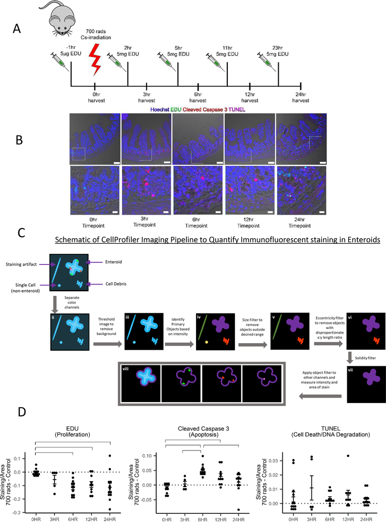
Figure 1: Enteroids as a model for the progression of death and proliferation in the jejunum.
(A) Diagram of low dose irradiation treatment time course of C57BL/6J mice.
(B) Immunofluorescence staining of 4% PFA fixed paraffin embedded 5μm sections from mice 0, 3, 6, 12, and 24 hours post 700 rad 137Cs-irradiation. Hoechst (nuclei), EDU (proliferating cells), Cleaved Caspase-3 (apoptosis), TUNEL (late stage apoptosis/necrosis). Imaged on a Zeiss LSM880 with a 20X air objective. Scale bar = 50 μm. Below are digitally zoomed in images of the crypts using photoshop, scale bar = 10 μm.
(C) Graphical representation of the Cell Profiler pipeline used to analyze 137Cs-irradiated enteroids.
(D) Quantification of fluorescence intensity of EDU, Cleaved Caspase-3, and TUNEL staining in 700 rad 137Cs-irradiated enteroids. Irradiated samples were normalized to non-irradiated samples and as a function of the size of the enteroid. Data acquired from 12 independent mouse preparations of enteroids on 3 separate days. Significance was determined by 2-way ANOVA with Tukey post-test (p < 0.05) and is indicated by the brackets. (See also Figure S1)

