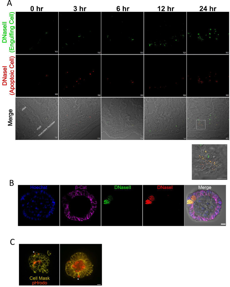
Figure 2: Paneth Cells can act as phagocytes within the small intestine.
(A) Immunofluorescence staining of 4% PFA fixed paraffin embedded 5μm sections from mice 0, 3, 6, 12, and 24 hours post 700 rad 137Cs-irradiation. Apoptag staining shows DNaseI (red) and DNaseII (green) staining. Imaged on a Zeiss LSM880 with a 63X oil objective. Scale bar = 10 μm. (See also Figure S2).
(B) Apoptag staining of 4 day old enteroids irradiated with 700 rads 137Cs-irradiated 6 hours prior to fixation with Cytofix/Cytoperm. Enteroids were also stained with nuclear counterstain, Hoechst (blue), and beta catenin (magenta). Imaged on a Zeiss LSM880 63X oil objective. Scale bar = 10 μm.
(C) Frame from live imaging of membrane labeled UV irradiated 4 day old enteroids (CellMask, yellow) co-stained with pHrodo. Asterix indicates engulfed cargo within an intact intestinal epithelial cell. Imaged on a Leica DMi8 inverted motorized microscope with temperature and CO2 controlled OKO chamber. Images were captured with a 63X oil objective. Scale bar = 10 μm. (See also Video S1 and Video S2)

