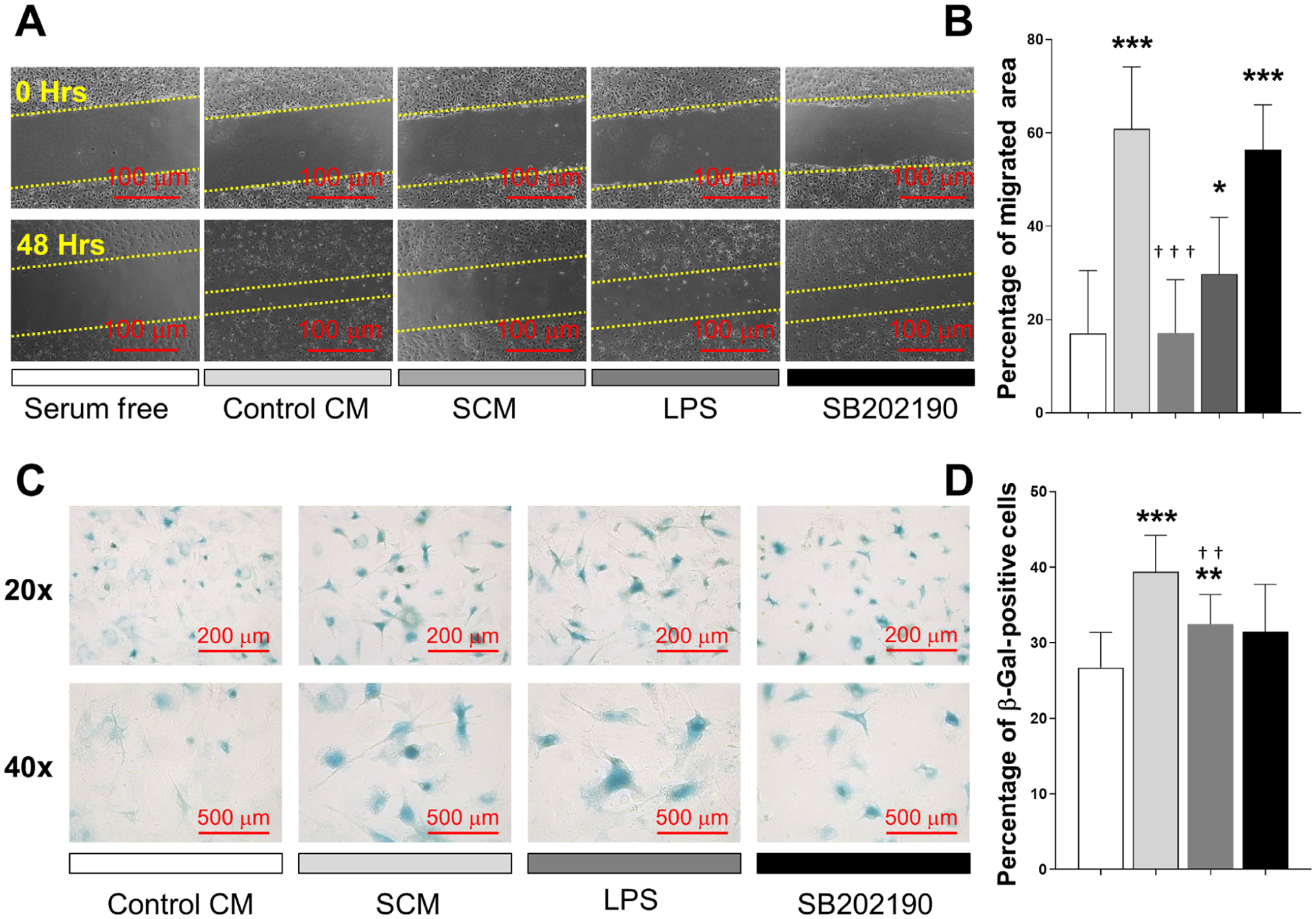
FIGURE 5.

Senescent osteocyte secreted factors induce a deleterious effect on osteoprogenitor cells. A and B) A wound healing assay was used to measure the effect of SCM on chemotactic cell recruitment. Cells were exposed to serum free media as control, CM from healthy osteocytes (control CM), SCM from irradiated osteocytes, LPS (10 μg/mL), or SCM plus SB202190 (10 μM). Cells were allowed to migrate into the scratched area for 48 hours. Representative images from each condition are displayed, and results presented as percentage of migrated area. C and D) The acquisition of senescence-associated SA-β-activity was evaluated by treating primary cells with control CM, SCM, LPS (10 μg/mL) or SCM combined with SB202190. Representative images from each condition are displayed and the results are presented as percentage of β-gal positive cells. Significant changes are indicated by: * compared with serum free or control CM, and † compared with control CM versus SCM or SCM versus SB202190. Various levels of significance are based on the number of each respective symbol (one symbol P < 0.05; two symbols P < 0.01; three symbols P < 0.001)
