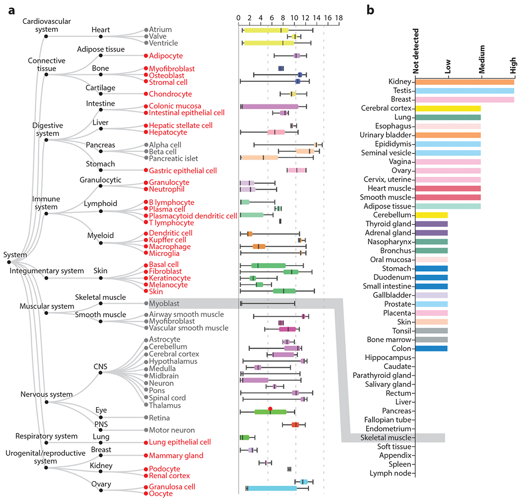
Figure 3.

Tissue expression pattern of CaV1.2. (a) Figure and data are adapted from the ARCHS4 web resource (https://amp.pharm.mssm.edu/archs4/gene/CACNA1C) showing CACNA1C expression in various tissues. In red are nonexcitable cell types with expression of CaV1.2. Note that expression levels for many of these cell types are like the expression level in the heart, the canonical CaV1.2-expressing tissue. In contrast, skeletal muscle myoblasts, which express an alternative L-type voltage-gated Ca2+ channel (CaV1.1), show no expression (highlighted by gray box). (b) Figure and data for protein detection (adapted from https://www.proteinatlas.org/ENSG00000151067-CACNA1C/tissue) (55) also demonstrate broad tissue expression, with no CaV1.2 protein detected in skeletal muscle (highlighted by gray box).
