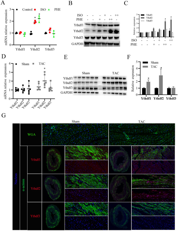
Fig. 2.
YTHDF2 protein expression increases in the heart tissues of mice with cardiac hypertrophy. A RT-PCR analysis of YTHDF1/2/3 mRNA expressions in the primary cardiomyocytes stimulated with isoproterenol (ISO, 10 μmol/l), phenylephrine (PHE, 50 μmol/l), or DMSO (as control) for 24 h. B Western blotting analysis of YTHDF1/2/3 protein expressions in the primary cardiomyocytes stimulated with ISO (+ , 10 μmol/l; + + , 20 μmol/l), PHE (+ , 50 μmol/l; + + , 80 μmol/l), or DMSO (as control) for 24 h. C Densitometry quantification of protein expressions. D RT-PCR analysis of YTHDF1/2/3 mRNA expressions in the heart tissues of mice after 4 weeks of TAC surgery (n = 6) or Sham surgery (n = 6). E Western blotting analysis of YTHDF1/2/3 protein expressions in the heart tissues of Sham (n = 6) or TAC (n = 6) mice. F Densitometry quantification of protein expressions. G WGA staining (Green) was used to measure cardiomyocyte size in the heart tissues of Sham or TAC mice; Immunofluorescence analysis of YTHDF1/2/3 (Red) expressions in cardiomyocytes (stained with anti-α-actinin antibodies, Green). Nucleus was stained with DAPI (Blue). *P < 0.05

