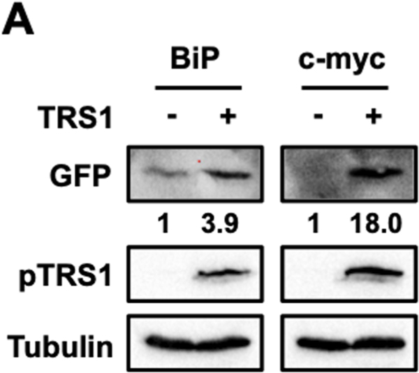
Figure 4. Cellular IRES activity is enhanced by pTRS1 expression.
HeLa cells were co-transfected with a circular GFP reporter containing either the BiP IRES (BiP) or the c-myc IRES (c-myc) with either a vector control or TRS1. A representative Western blot image showing GFP and pTRS1 (n=3). Numbers indicate the fold change in GFP expression compared to vector control, normalized to pTRS1 and tubulin levels.

