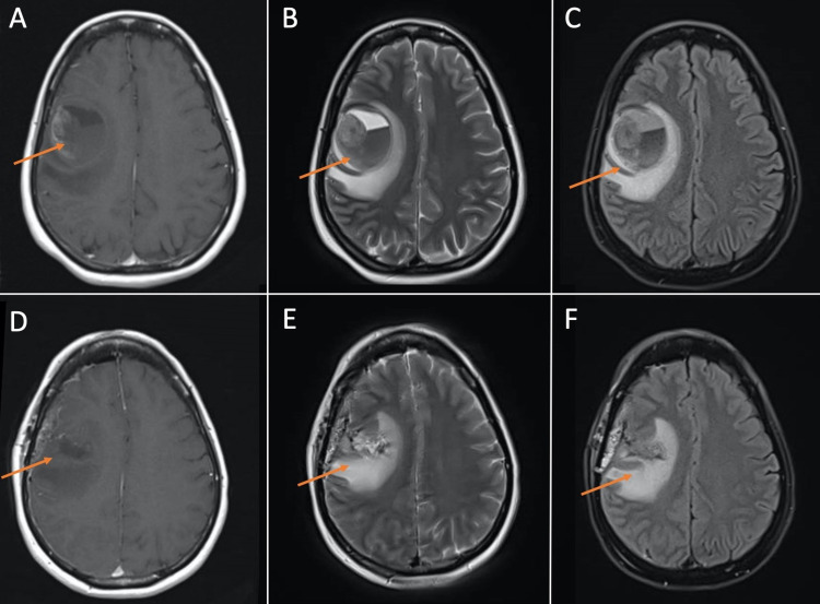
Figure 1. MRI brain demonstrating large hemorrhagic cavity in the right frontal lobe with an enhancing focus along the right superolateral margin representing a hemorrhagic mass. (A), (B), and (C) are pre-operative T1, T2, and FLAIR images, respectively. (D), (E), and (F) are post-operative T1, T2, and FLAIR images, respectively, demonstrating resection of mass and post-surgical changes.
FLAIR: fluid-attenuated inversion recovery.

