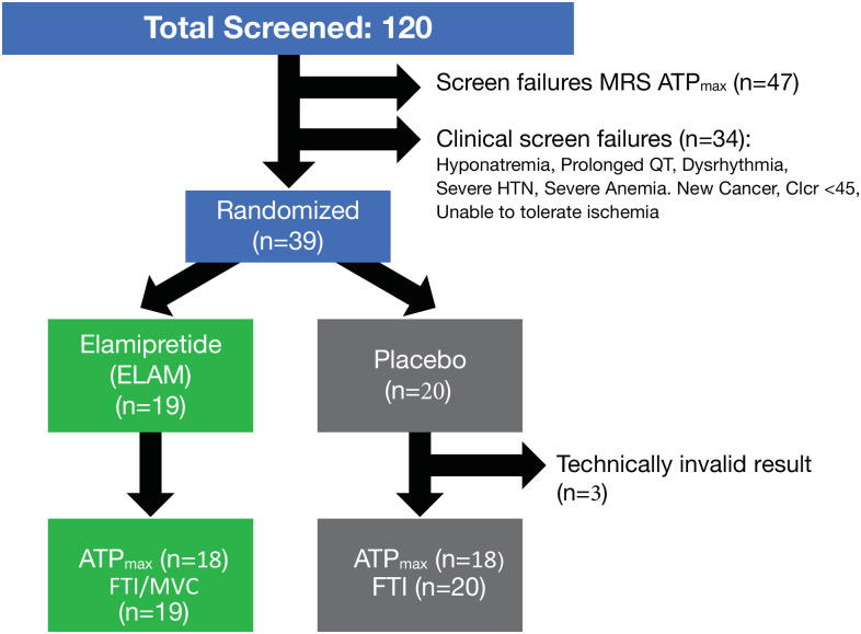
Fig 1. Study schema.
The primary endpoint for the study was change in ATPmax by phosphorus magnetic resonance spectroscopy (31P MRS) after a 2-hour infusion compared with baseline (screening). The secondary endpoints were a change in mitochondrial coupling (P/O) and muscle force time integral (FTI) compared with baseline (screening).

