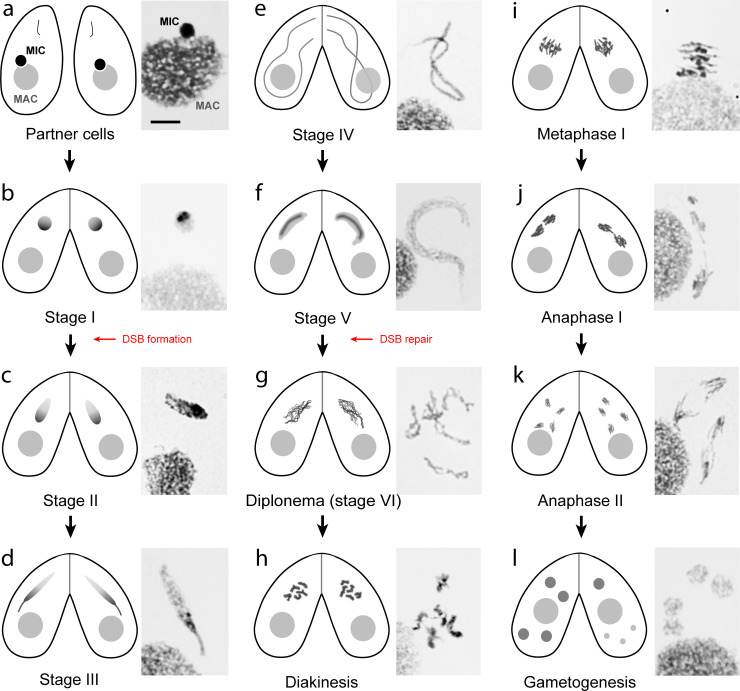
Fig 2. Tetrahymena meiotic stages.
Schematic diagrams of mating cells and microscopy images of Giemsa-stained meiotic nuclei. Meiotic prophase is staged according to [79]. (a) In nonmeiotic cells, the MIC is located in a pocket on the MAC surface. (b) In stage I conjugating cells, the round MIC moves away from the MAC. The progress of meioses in conjugated cells is largely synchronous. (c–e) Once DSBs begin to form, the MICs start stretching and elongate to about twice the length of the cell. (f) MICs then shorten, and all DSBs are repaired by the end of stage V. (g) Progressive chromatin compaction reveals the formation of 5 bivalents. (h) Bivalents with protruding centromeres have reached maximal condensation. (i) Bivalents arranged in a metaphase plate. (j, k) Anaphase I and II follow the conventional scheme. (l) After telophase II (left cell), one of the 4 meiotic products is selected to divide into 2 gametic nuclei, whereas the other 3 products degenerate (right cell). Bar: 10 μm. DSB, double-strand break; MAC, macronucleus; MIC, micronucleus.

