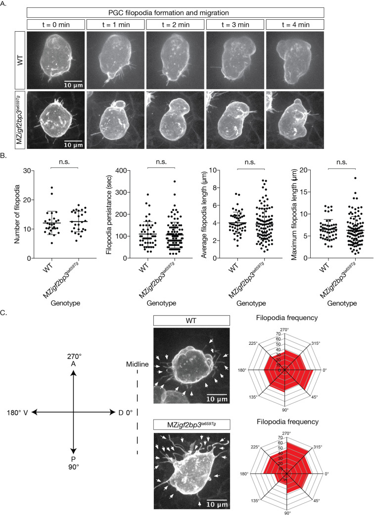
Fig 6. PGC membrane projections show altered behaviour in MZigf2bp3la659Tg mutants.
A. Images of cell membranes of PGCs labelled with a Farnesylated-GFP-nos1 live reporter in bud stage mutant or wild type embryos. B. Quantitation of filopodia and behaviour (persistence, length) in WT and MZigf2bp3la659Tg PGCs. C. Projection angle relative to their destination, showing an altered skew in maternal igf2bp3la659Tg PGCs, although the projection parameters, namely numbers, persistence, average and maximum lengths did not appear to be changed. p *<0.05, **<0.01, ***<0.001. N = 27 WT and 26 MZigf2bp3la659Tg PGCs; number of filopodia = 50 WT and 100 mutant filopodia. Number of PGCs analysed for projection frequency = 25 per genotype; Scale bars, 10 μm.

