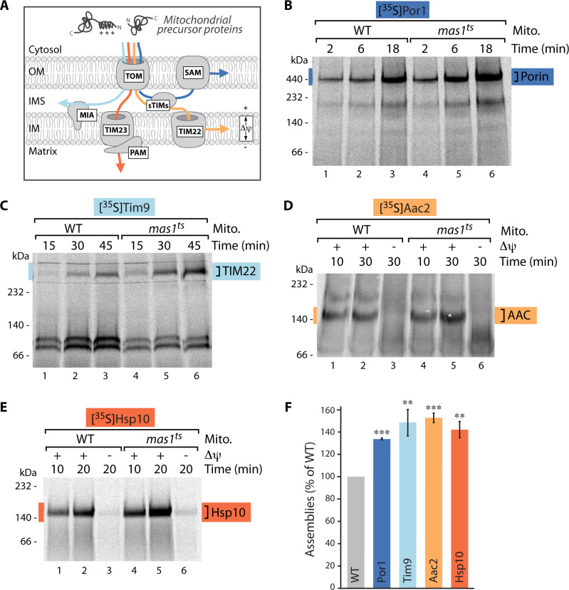
Fig 1. Protein import into all mitochondrial subcompartments is stimulated upon mtUPR.
(A) Schematic of the main protein import pathways into mitochondria. Protein translocation into the matrix and inner membrane depends on the membrane potential (Δψ) as driving force, protein import into the outer membrane or intermembrane space is membrane potential-independent. TOM, translocase of the outer membrane; SAM, sorting and assembly machinery; sTIMs, small TIMs; MIA, mitochondrial intermembrane space assembly; TIM23 and TIM22, translocases of the inner membrane; PAM, presequence translocase associated motor; OM, outer mitochondrial membrane; IMS, intermembrane space; IM, inner mitochondrial membrane. (B)-(E) Import kinetics of indicated radiolabeled precursor proteins into mitochondria isolated from wild-type (WT) or mas1ts cells after induction of mtUPR for 10 hours. Where indicated (in case of matrix- and inner membrane-targeted substrates), the membrane potential (Δψ) was dissipated prior to the import reaction. Samples were solubilized in the mild detergent digitonin and analyzed by BN-PAGE and autoradiography. In Fig 1C the soluble Tim9/Tim10 complex is formed at appr. 100 kDa. (F) Quantification of longest import time-point displayed in (B)-(E) normalized to WT value. n = 3, data represent means ± SEM. Student’s t-test was used for pairwise comparison. **p < 0.01; ***p < 0.001.

