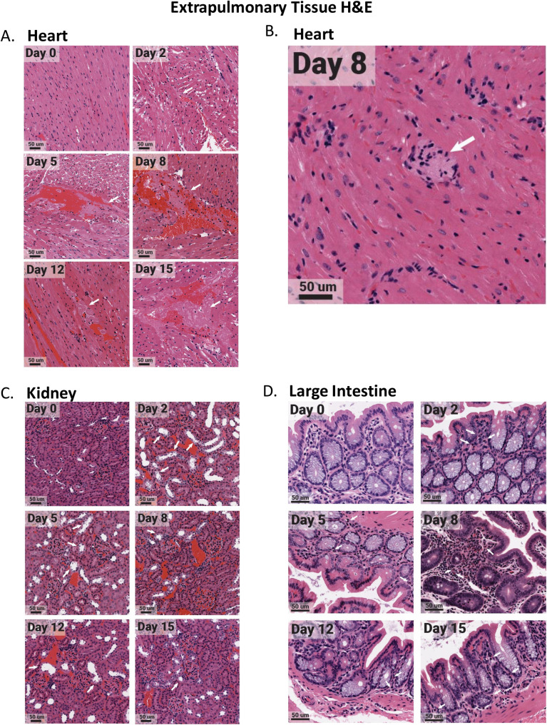
Fig 7. Select extrapulmonary organs have distinct pathology after SARS-CoV-2 infection with evidence of microthrombi and myocarditis in the heart, tubular damaging in the kidney, and eosinophil infiltration in the large intestine.
The heart, kidney, and large intestine were collected from each animal at necropsy and fixed in 10% formalin prior to paraffin block embedding. Tissues were H&E stained. Heart tissues exhibited microthrombi (white arrows) over multiple days (A), as well as myocarditis on day 8 (white arrow) (B). The kidney had acute injury/tubular injury (white arrows) (C). The large intestine had eosinophil infiltration (white arrows) (D). Stained tissues were visualized and imaged using the Aperio ScanScope XT slide scanner. Images shown are representative of 3 animals per group, per timepoint.

