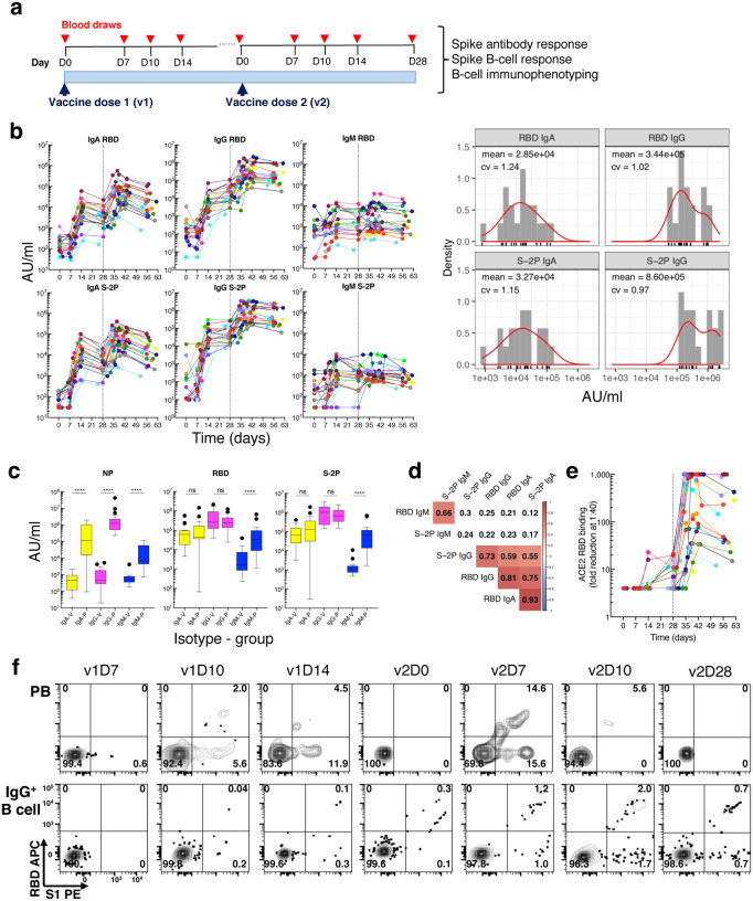
Fig. 1. Longitudinal blood sampling and analysis shows robust antibody and early B cell response to mRNA-1273 vaccine.
a, Study design with serial blood draws and assays performed at all timepoints on SARS-CoV-2-uninfected vaccinees (n = 21; missed visits in Extended Data Table 1) receiving two doses of the mRNA-1273 vaccine. b, Serum IgG, IgA and IgM binding to S-2P and RBD proteins measured by electrochemiluminescence (ECLIA) longitudinally (left panels), and corresponding histogram and distribution (based on kernel density estimates) at the last timepoint (v2D28) (right panels). c, Peak serum IgG, IgA and IgM binding to S-2P, RBD and N proteins measured by ECLIA in vaccinees (V; n = 21) and COVID-19 patients (P; n = 21), shown as boxplots. d, Triangular heatmap of correlation between serum antibodies at last measured timepoint (v2D28) in (b). Numbers represent r values. Statistically insignificant correlations (p > 0.05) shown in white. e, Longitudinal inhibition of RBD binding to ACE2 by serum (1:40 dilution) of vaccinees (n = 21). f, Longitudinal binding of S1 and RBD tetramers to PB and IgG+ B cells by flow cytometry shown for a high responder (VAC-611; Extended Data Table 1). Numbers in each quadrant are percentages. Each vaccinee is color-coded and second vaccine dose indicated by vertical dotted line (b,e). Mann-Whitney test; ****, p < 0.0001 (c). Spearman’s rank correlation (d). AU, arbitrary units; D, day; N, nucleocapsid; ns, not significant; P, patients with severe COVID-19; PB, plasmablasts; RBD, receptor binding domain; S1, spike subunit 1; S-2P, stabilized spike trimer; v, vaccine dose; V, vaccinees.

