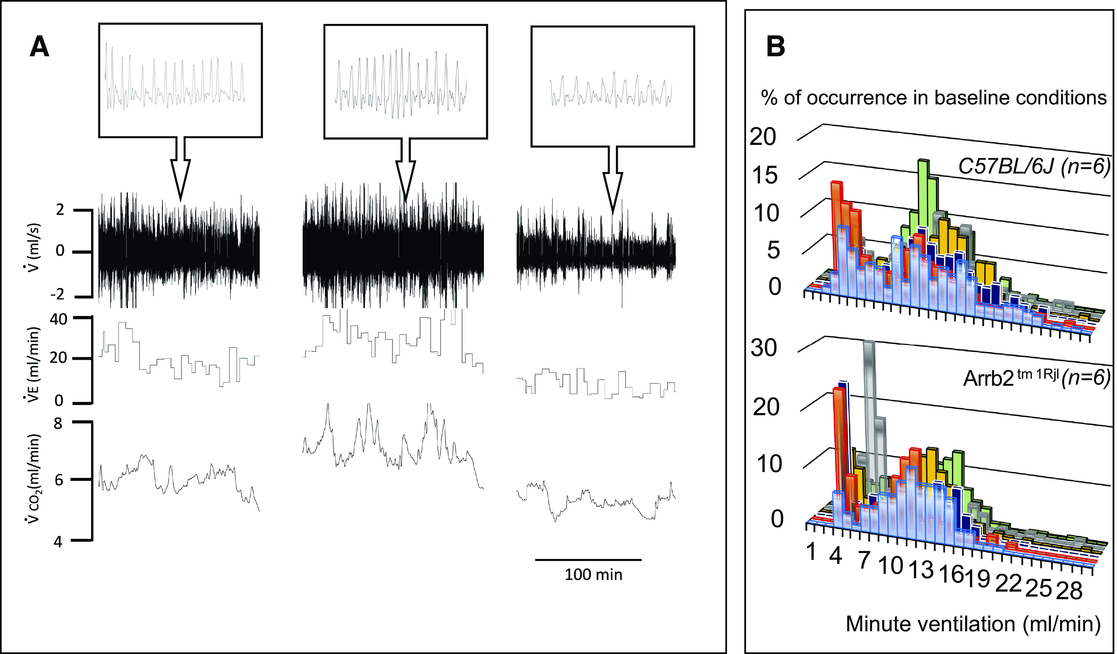
Figure 2.

A: example of a recording of baseline spontaneous breathing in one control mouse. The mouse was exposed to dim light and placed in a plethysmographic box for 2 h. Spontaneous instantaneous respiratory flow (V̇), minute ventilation (V̇e), and V̇co2 are shown. Note the spontaneous change in breathing associated with proportional changes in metabolic rate, reflecting fluctuations in the level of vigilance, in behavior and motor activity. B: frequency distribution of ventilation during a 1 h-period of recording in all control (top) and β-arr2-deficient (bottom) mice. Note the first peak corresponds to the lowest levels of ventilation measured when animals were immobile. Arrb2tm1Rjl, Arrb2 knockout mice; β-arr2, β arrestin 2.
