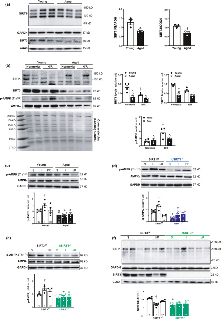
FIGURE 1.

Age‐related decline of SIRT1 and SIRT3 causes the impairment of AMPK activation during myocardial I/R. (a) The protein expression level of SIRT1 and SIRT3 was decline in the left ventricle of aged (24–26 months) C57BL/6J hearts. Values are mean ± SEM. N = 3, *p < 0.05 vs. young (4–6 months) C57BL/6J hearts. (b) Expression of SIRT1 and SIRT3 in protein level was reduced with aging in the isolated cardiomyocytes. Their protein levels were decline in response to H/R in the cardiomyocytes of young hearts, and this stress‐related response was blunted in aged hearts. Values are mean ± SEM. N = 4, *p < 0.05 vs. young; † p < 0.05 vs. normoxia. (c) 30‐min ischemia and 6‐h reperfusion could activate AMPK, and the AMPK phosphorylation was impaired in aged hearts during I/R. Values are mean ± SEM. N = 4, *p < 0.05 vs. young; † p < 0.05 vs. sham. (d) The AMPK phosphorylation induced by cardiac I/R was blunted in the left ventricle of icSIRT1−/− (4–6 months) hearts. Values are mean ± SEM. N = 4, *p < 0.05 vs. SIRT1 f/f (4–6 months); † p<0.05 vs. sham. (e) The activation of AMPK during myocardial I/R was impaired in the left ventricle of cSIRT3−/− (4–6 months) hearts. Values are mean ± SEM. N = 4, *p<0.05 vs. SIRT3 f/f (4–6 months); † p<0.05 vs. sham. (f) The protein level of SIRT1 was decreased during myocardial ischemia. Cardiomyocyte‐specific deletion of SIRT3 caused the reduction of SIRT1 expression in the left ventricle of cSIRT3−/− hearts. Values are mean ± SEM. N = 4, *p<0.05 vs. SIRT3 f/f ; † p<0.05 vs. sham.
