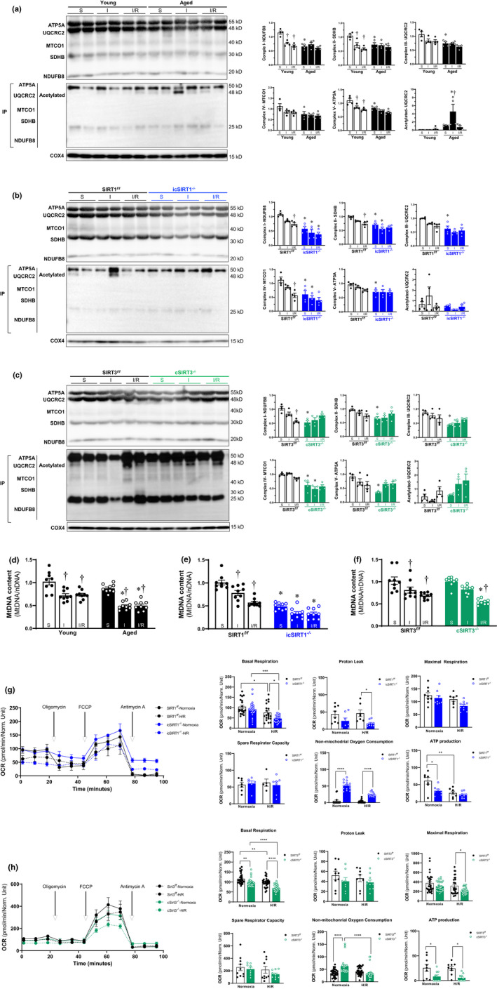
FIGURE 3.

Deficiency of SIRT1 and SIRT3 in aging impaired the integrity of OXPHOS complexes under stress condition. (a‐c) Immunoprecipitation and immunoblotting analysis of OXPHOS complex I, II, III, IV, V subunits accumulation levels and acetylation in young (4–6 months)/aged (24–26 months) WT (a), SIRT1 f/f /icSIRT1−/− (4–6 months) (b), SIRT3 f/f /cSIRT3−/− (4–6 months) (c) hearts under physiological and I/R stress conditions. Values are mean ± SEM. N = 4, *p < 0.05 vs. young, SIRT1 f/f , SIRT3 f/f , respectively; † p < 0.05 vs. sham, respectively. (d‐f) Real‐time PCR measured the relative mitochondrial DNA (mtDNA) content (normalized to the nuclear gene) in the heart tissue of young/aged WT (d), SIRT1 f/f /icSIRT1−/− (e), SIRT3 f/f /cSIRT3−/− (f) hearts. Values are means ± SEM, n = 9, † p < 0.05 vs. sham, respectively; *p < 0.05 vs. young, SIRT1 f/f , SIRT3 f/f , respectively; (g‐h) Mitochondrial respiration measurements of OCR in isolated cardiomyocytes of SIRT1 f/f /icSIRT1−/− (g), SIRT3 f/f /cSIRT3−/− (h) hearts were performed with a Seahorse metabolic analyzer. Oligomycin (1.5 μM), FCCP (1 μM), and antimycin (10 μM) were added sequentially to isolated cardiomyocytes under normoxia and H/R conditions. Quantitative analysis of mitochondrial function parameters (basal respiration, proton leak, maximal respiration, spare respiratory capacity, non‐mitochondrial oxygen consumption, and ATP production) is shown in the bar charts. Values are mean ± SEM, n ≥ 5, *p < 0.05 vs. SIRT1 f/f , SIRT3 f/f , respectively; † p < 0.05 vs. normoxia, respectively
