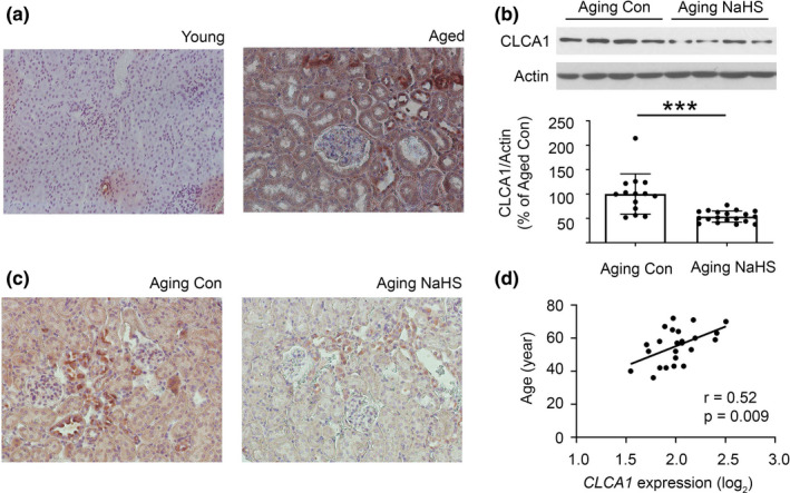
FIGURE 2.

Histologic analysis of kidney CLCA1 expression. (a) On immunoperoxidase staining (x200) a faint tubular expression of CLCA1 was seen in kidney cortex from young mice, which was robustly increased in aged mice. Representative images from young (n = 4 ) and aged mice are shown (n = 4). (b, c) Administration of NaHS to 18–19‐month‐old aging mice daily for 5 months (n = 20; Aging NaHS) reduced renal cortical CLCLA1 expression by immunoblotting compared to aging mice receiving water vehicle (Aging Con; n = 14; b). This was confirmed by immunoperoxidase staining (x200; C). (d) There was a direct correlation between age and CLCA1 mRNA expression in the tubule interstitium compartment in the kidney tissue from human subjects (n = 24). (b) Data (mean ± SD) are shown in bars with scatter plots and were analyzed by t test. ***p < 0.001
