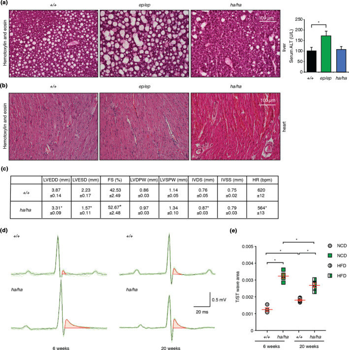
FIGURE 2.

Mrps12ha / ha mice develop hypertrophic cardiomyopathy on a high‐fat diet and Mrps12ep / ep mice have increased liver lipid accumulation. (a) Liver sections from Mrps12 +/+, Mrps12ep / ep and Mrps12ha / ha mice fed a high‐fat diet were cut to 10 µm thickness and heart sections (b) were cut to 5 µm thickness, then stained with haematoxylin and eosin (H&E). Each image is representative of sections from six mice per genotype. (c) Parameters for Mrps12 +/+ n = 5 and Mrps12ha / ha n = 5 mice fed a HFD. LVEDD, left ventricular end‐diastolic diameter; LVESD, left ventricular end‐systolic diameter; FS, fractional shortening; LVDPW, left ventricular posterior wall in diastole; LVSPW, left ventricular posterior wall in systole; IVDS, intraventricular septum in diastole; IVSS, intraventricular septum in systole; HR, heart rate. Values are means ± SEM. *p < 0.05 compared with Mrps12 +/+, Student's t test. (d) Representative raw electrocardiographic recordings from at least six control and six Mrps12ha / ha mice fed an NCD at 6 weeks and fed a high‐fat diet at 20 weeks. Characteristic altered repolarisation in hearts of Mrps12ha / ha mice was identified as an increase in the T/ST‐wave area. (e) Electrocardiography recordings showed significant differences in the T/ST‐wave area of the Mrps12ha / ha compared to Mrps12 +/+ mice fed either a normal diet at six weeks of age or following a high‐fat diet by 20 weeks of age. Values are means ± SEM. *p < 0.05 compared with Mrps12 +/+, Student's t test
