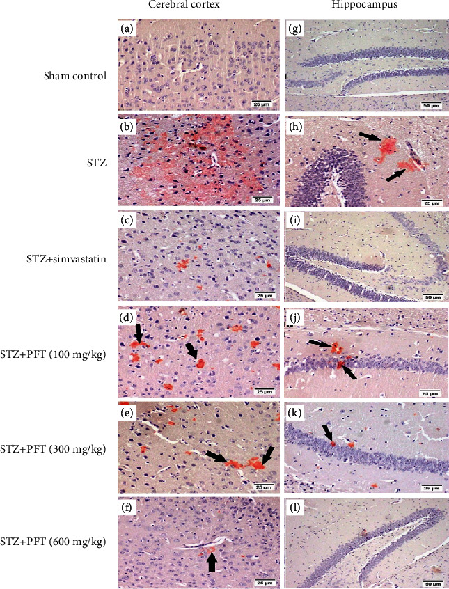
Figure 10.

Congo red stained brain sections of mice for amyloid plaques visualization. Sham control group (a, g) showed no deposition of amyloid plaques (magnification ×20 and ×10, respectively). STZ group (b, h) showed diffuse deposition in the cerebral cortex and multifocal deposition in the hippocampus (arrows), respectively (magnification ×20). STZ + simvastatin (c, i) displayed minute deposition of amyloid plaques (magnification ×20 and ×10, respectively). PFT (100 mg/kg) (d, j) revealed multifocal scattered plaques (arrows). PFT (300 mg/kg) (e, k) presented multifocal area in the cerebral cortex and few depositions in the hippocampus (magnification ×20). PFT (600 mg/kg) (f, l) exhibited minute deposition of amyloid plaques (magnification ×20 and ×10, respectively).
