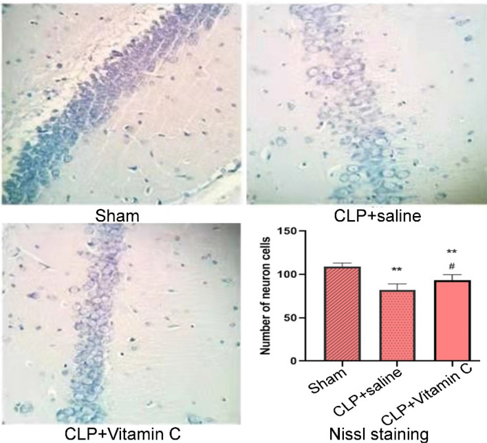
Figure 2.
Nissl staining in the hippocampal CA1 region. Representative sections from the hippocampal CA1 region were observed 24 h after operation (Nissl staining × 200). The pyramidal neurons in the hippocampal CA1 region were significantly decreased (P = 0.000) in both the CLP + saline group and the CLP + vitamin C group compared with the sham group. Compared with the CLP + high-dose vitamin C group, the pyramidal neurons in the CLP + saline group were significantly (P = 0.01) decreased. *P < 0.05 and **P < 0.01 compared with sham group; #P < 0.05 compared with the CLP group. CLP, cecal ligation and puncture. Statistical value: 24.69, total degree of freedom: 14, P: 0.000.

