Fig. 4.
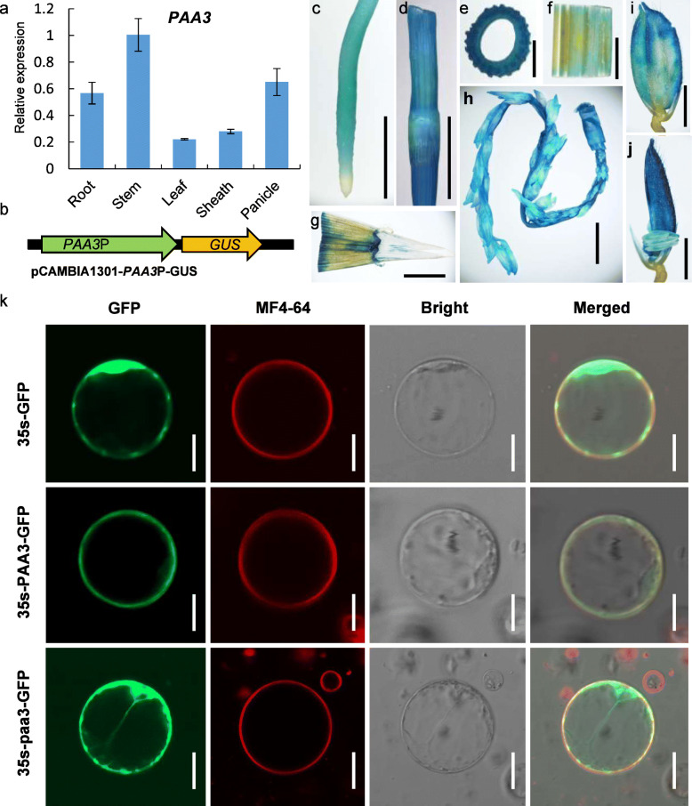
Expression pattern analysis of the PAA3 gene and subcellular localization of PAA3 protein. a: qPCR analysis of rice paa3 from different tissues of WT normalized to actin. b: Structure of PAA3P-GUS. c–j: Tissue-specific expression of the GUS gene driven by the rice paa3 promoter. Root (c) Stem (d, e) Leaf blade (f) Sheath (g) Panicle (h), Spikelet (i) Spikelet without lemma (j). k: Analysis of the subcellular localization of the PAA3 protein. 35 s-GFP indicates the expression of GFP protein without PAA3 in rice protoplasts as the negative control. 35 s-PAA3-GFP indicates the plasma membrane localization of the PAA3 protein in rice protoplasts given by the expression of PAA3 fused with GFP. 35 s-paa3-GFP indicates the cytoplasm localization of the paa3 protein in rice protoplasts given by the expression of paa3 fused with GFP. The paa3 protein is the mutant protein. Green is GFP signal. Red is the plasma membrane signal, which was labelled by MF4–64. Bright is the bright light. Merged indicates the confused with the Green signal, the Red signal, and the bright light. Bars: (c–h) 10 mm; (I and j) 1 mm; (k) 50 μm

