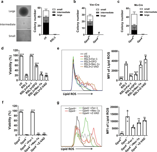
Fig. 1. GPX4 deficiency induces HSPC ferroptosis in vitro.
a LT-HSC single cells were sorted and cultured in a medium with or without RSL3 for 14 days. Colony numbers were counted (n = 4 mice). Scale bars = 0.5 mm. LT-HSCs derived from the Gpx4flox/flox mice and Gpx4flox/flox Vav-Cre mice (b) or the pIpC-treated Gpx4flox/flox Mx-Cre mice (c) were tested with single-cell colony-forming assay as in (a) (n = 2 Vav-Cre mice or 4 Mx-Cre mice). d The viability of LSK cells isolated from the wild-type mice after 48 h of culture with the indicated drugs (n = 3 mice). e LSK cells isolated from the wild-type mice were cultured with the indicated drugs for 24 h, and lipid ROS were measured with C11-BODIPY and detected by flow cytometry (n = 3 mice). f The viability of LSK cells isolated from the Gpx4flox/flox mice or the Gpx4flox/flox Vav-Cre mice after 48 h of culture with the indicated drugs (n = 3 mice). g LSK cells isolated from the Gpx4flox/flox mice or the Gpx4flox/flox Vav-Cre mice were cultured with the indicated drugs for 24 h, and lipid ROS were measured with C11-BODIPY and detected by flow cytometry (n = 3 mice). Data are the mean ± SD. (ns not significant, *P < 0.05, **P < 0.01, ***P < 0.001 vs. the RSL3 group in (d, e), vs. the Gpx4−/− group in (f, g).

