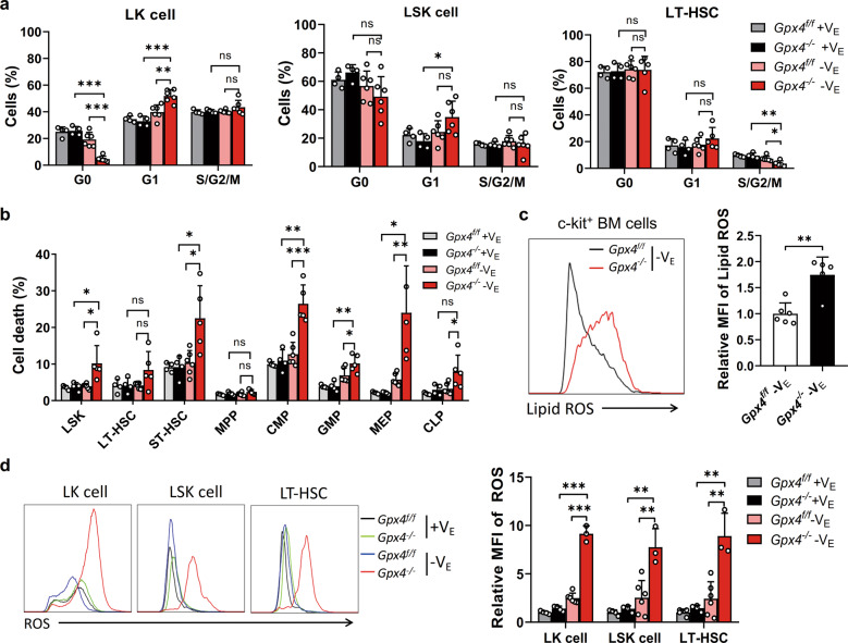
Fig. 6. Deficiency of both GPX4 and vitamin E results in HSC ferroptosis in vivo.
a The cell cycle of LK cells, LSK cells, and LT-HSCs in the Gpx4flox/flox Vav-Cre mice after 3 weeks of a VE-depleted diet. b The HSPCs in the Gpx4flox/flox Vav-Cre mice after 3 weeks of a VE-depleted diet were determined to be DAPI negative. c Left panel: representative flow cytometric plots of lipid ROS in c-kit+ cells of the Gpx4flox/flox Vav-Cre mice after 3 weeks of a VE-depleted diet. Right panel: statistical data of the relative MFI of lipid ROS. d Left panel: representative flow cytometric plots of ROS in LK cells, LSK cells, and LT-HSCs of the Gpx4flox/flox Vav-Cre mice after 3 weeks of a VE-depleted diet. Right panel: statistical data of the relative MFI of ROS. N = at least three mice in each group. Data are the mean ± SD. (ns not significant, *P < 0.05, **P < 0.01, ***P < 0.001).

