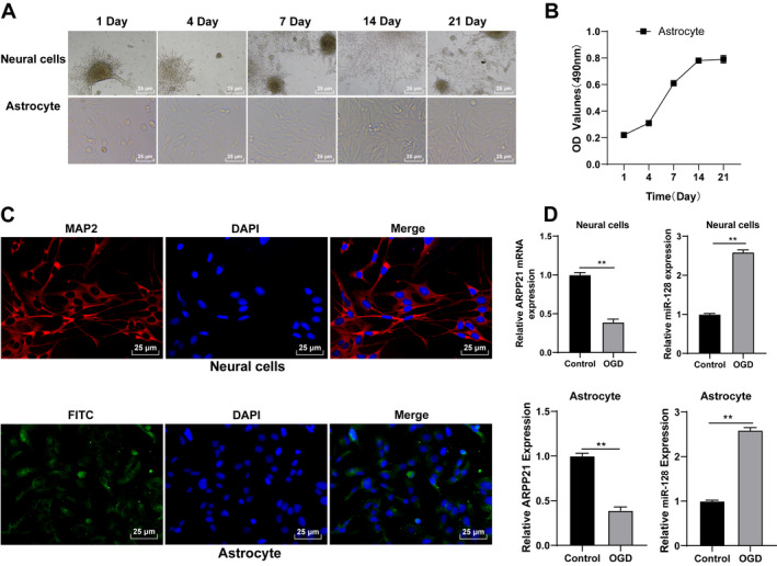
FIGURE 2.

Astrocytes were incubated and identified in vitro. (A) Morphological changes in neurons and astrocytes were observed under the inverted phase‐contrast microscope. (B) Proliferation ability of astrocytes was detected using MTT assay. (C) Neurons and astrocytes were identified using immunofluorescence. (D) Expressions of ARPP21 and miR‐128 in cells were detected using RT‐qPCR. The cell experiment was repeated three times independently. Data were expressed as mean ± standard deviation. Data in panel (B/D) were analyzed using t test, **p < 0.01.
