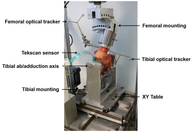
Figure 1.

Experimental setup of a left knee at 9° lateral downslope and 20° of knee flexion. The femoral pot was secured to the fixture mounted on the materials testing machine actuator at the top of the picture. The tibial rig sits on the XY rolling table, which allowed free movement including free abduction and adduction about the AP pivot axis. Optical trackers were securely fixed to the femur and tibia to measure relative movements of the femur and tibia while axial force was applied. A foil pressure sensor was inserted underneath the medial and lateral menisci to obtain joint pressure readings.
