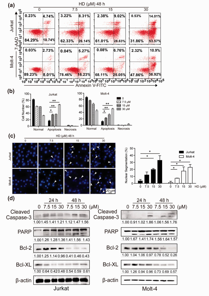
Figure 3.
HD induces apoptosis in T-ALL cells. (a) Annexin V/7-AAD double staining and flow cytometry analysis were used to detect the apoptosis ratio of cells treated with HD (0, 7.5, 15, 30 µM) for 48 h. (b) Quantification of the number of apoptotic cells in panel A. HD vs. Control. *p< 0.05, **p< 0.01. (c) After DAPI staining, the nucleus morphology of T-ALL cells treated with HD was imaged and the number of apoptotic cells was quantified. Scale bar, 20 μm. (d) After HD treatment for 24 or 48 h, Western blotting analysis of apoptosis-related proteins was performed. (A color version of this figure is available in the online journal.)

