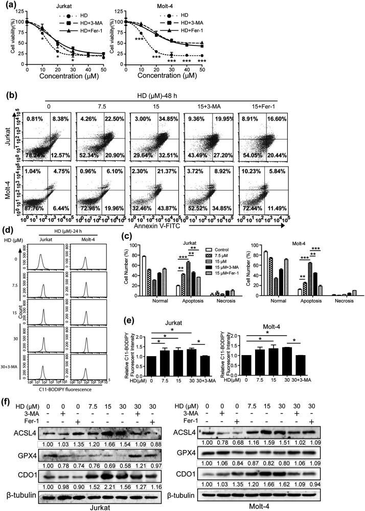
Figure 6.
Inhibition of autophagy reduces HD-induced ferroptosis in T-ALL cells. (a) After treated with HD in the present or absent of autophagy inhibitor 3-MA (2 mM) or ferroptosis inhibitor Fer-1 (10 µM), cell viability was determined by MTT assay. (b) After pre-treatment with either 3-MA (2 mM) or Fer-1 (10 µM) in combination with HD, cell apoptosis ratio was detected by Annexin V/7-AAD double staining and subsequent flow cytometry analysis. (c) Quantification of the number of apoptotic cells in panel b. **p < 0.01, ***p < 0.001. Lipid ROS production after treatment with HD with or without 3-MA (2 mM) for 24 hours was assessed by flow cytometry (d) and quantified in panel e using the C11-BODIPY probe. *p < 0.05. (f) Western blotting analysis of ferroptosis-related proteins in T-ALL cells after pre-treatment with 3-MA (2 mM) or Fer-1 (10 µM) for 2 hours, and then incubated with HD for another 24 h.

