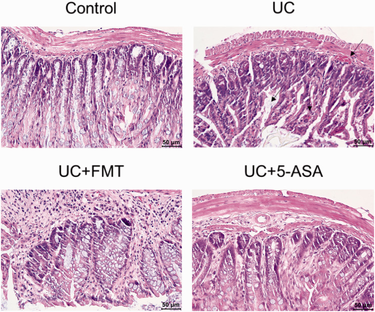
Figure 1.
HE staining of the intestinal tissue of mice in each group. For the model group, the mice that tested positive for fecal occult-blood screening were selected for verification by HE staining. A comparison between the UC and control groups was made for model verification, followed by a comparison between groups (UC, control, and the treatment groups UC+FMT and UC + 5-ASA). Note: scale 50 μm. Control: normal control, UC: ulcerative colitis model, UC+FMT: ulcerative colitis model + fecal microbiota transplantation, UC + 5-ASA: ulcerative colitis model + 5-aminosalicylic acid. (A color version of this figure is available in the online journal.)
HE: hematoxylin-eosin.

