Figure 3.
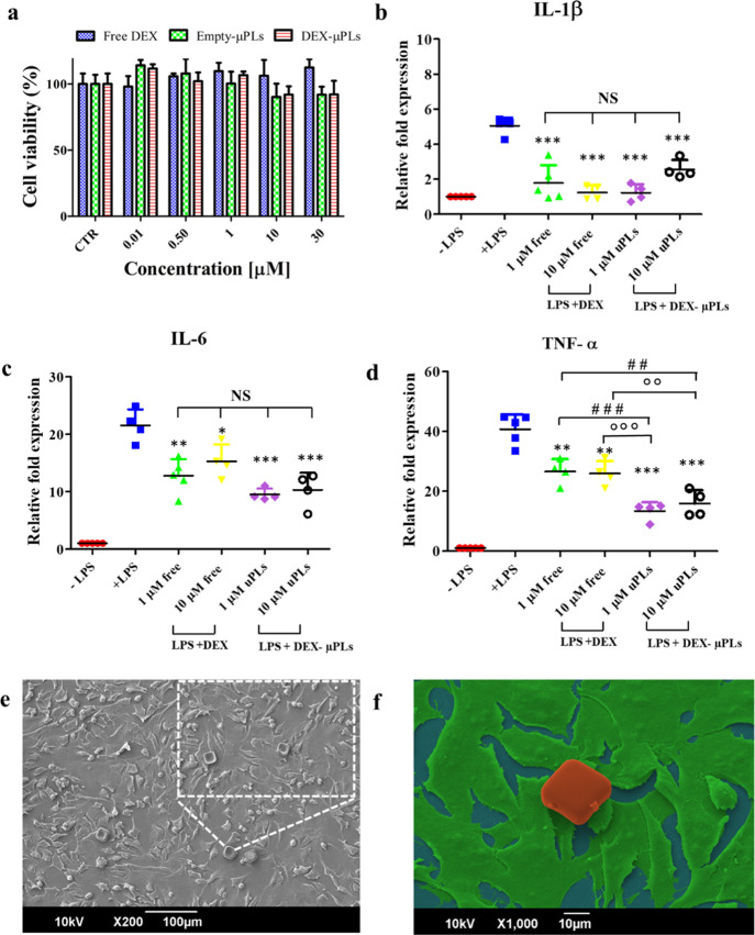
In vitro cytocompatibility and anti-inflammatory effect of DEX-loaded microPlates (DEX-μPLs). (a) ATDC5 cell viability upon incubation with free DEX, empty-μPLs, and DEX-μPLs. Statistical analysis via one-way ANOVA (GraphPad Prism 5) is provided in Table S3; (b–d) Expression levels of proinflammatory cytokines IL-1β, IL-6, and TNF-α for LPS-stimulated ATDC5 cells. (−LPS: no LPS and no μPLs; +LPS: LPS stimulation and no μPLs; DEX: LPS stimulation and free DEX at 1 and 10 μM; and DEX-μPLs: LPS treatment and DEX-μPLs at 1 and 10 μM). Results are presented as average ±SD (n ≥ 4). *p < 0.05, **p < 0.001, and ***p < 0.0001 were considered statistically significant as compared to the control (+LPS); ##p < 0.001 and ###p < 0.0001 were considered statistically significant as compared to 1 μM DEX; and oop < 0.001 and ooop < 0.0001 were considered statistically significant as compared to 10 μM DEX. The lack of a statistically significant difference between groups is indicated on the graphs as NS. Multiple comparisons were performed using, as the post hoc test, the Tukey’s significant difference (HSD) test; (e) 30° tilted view of a SEM image of ATDC5 cells incubated with μPLs. In the lateral inset, a magnified image shows cells interacting with μPLs; (f) False-color SEM image of a μPL (red) deposited and not internalized over a layer of ATDC5 cells (green).

