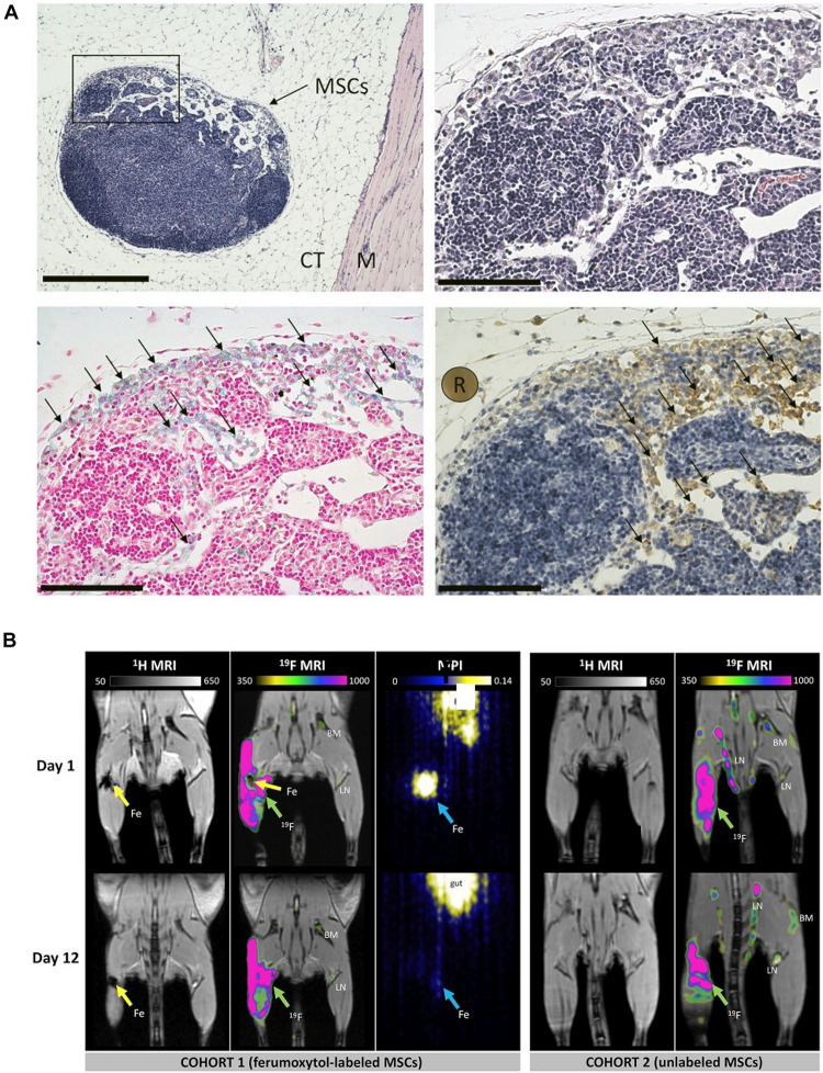
FIGURE 1.
(A) Histological validation showing the presence of MSCs surrounded in connective tissue (CT) and muscle (M) in hematoxylin and eosin at × 10 magnification (scale bar 500 μm). (B) In vivo proton (1H)/fluorine 19 (19F) magnetic resonance imaging (MRI) and magnetic particle imaging (MPI) (adapted Sehl et al., 2019).

