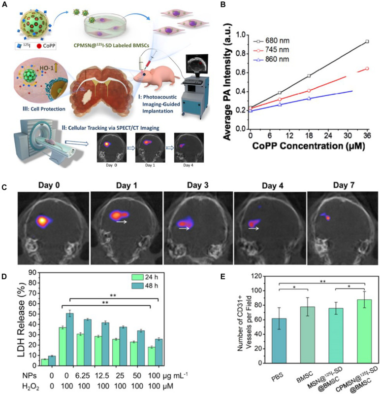
FIGURE 6.
(A) Schematic illustrating the triple functionality of CPMSN@125I-SD for stem cell therapy of brain ischemia. (B) Corresponding average intensities of CPMSN@125I-SD with various cobalt protoporphyrin IX (CoPP) concentrations in the NIR region. (C) Single-photon emission computed tomography imaging (SPECT)/CT images of ischemic mouse brain tissue on different days (0–7 days) after intracerebral injection of the labeled BMSCs (500,000 cells). (D) Cell death assessment for MSCs after treatment with different concentrations of CPMSN@125I-SD (0–100 μg ml–1) and exposure to 100 μM H2O2 for 24 and 48 h. (E) Bar graph showing the quantification of the number of CD31+ cells (adapted from Yao et al., 2020). ∗p < 0.05; ∗∗p < 0.01.

