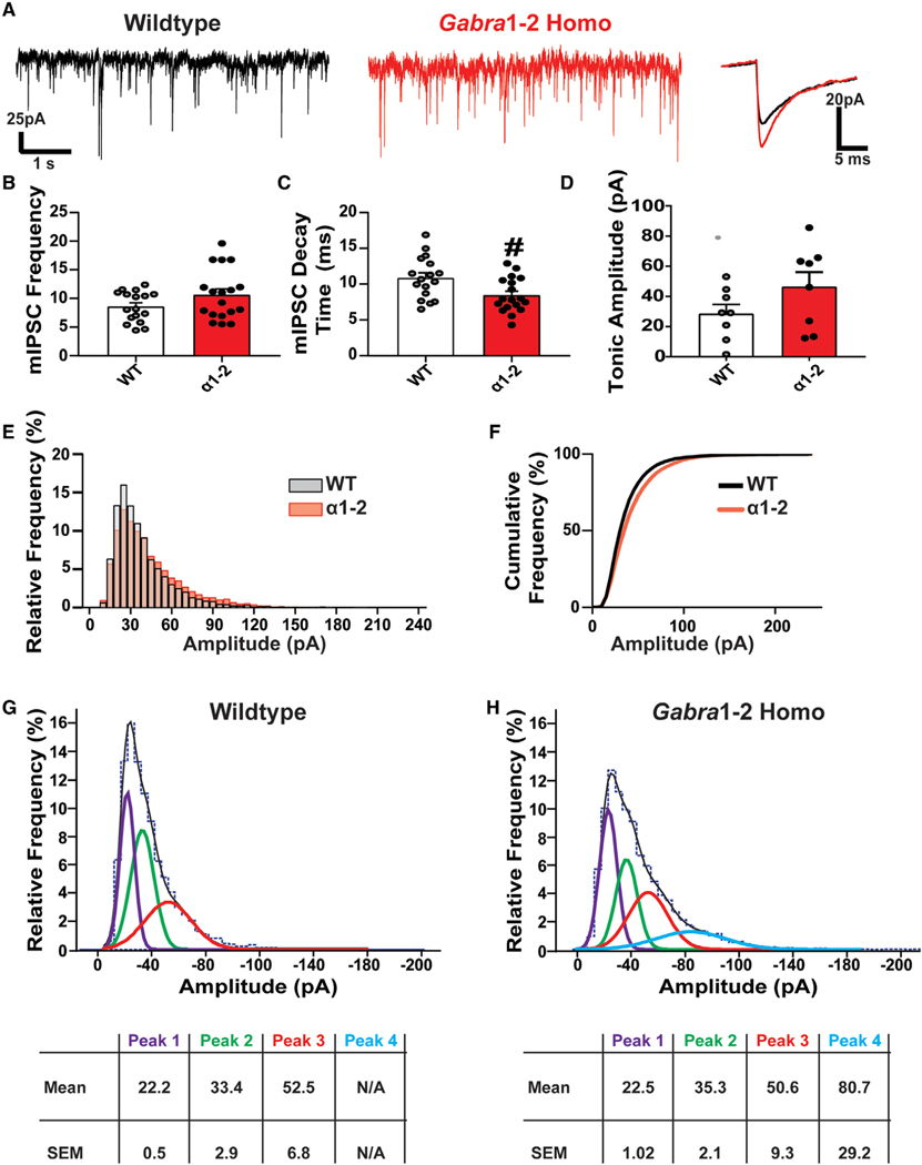
Figure 4. Miniature Inhibitory Postsynaptic Currents (mIPSC) Kinetics Are Altered in the Gabra1–2 Hippocampus.
(A) Representative mIPSC recordings from pyramidal neurons in the CΑ1 of WT and Gabra1–2 hippocampal slices, with superimposed spikes (right) representing the average WT (black) and Gabra1–2 traces (red).
(B and C) Quantification of mIPSC kinetics (n = 6 mice/genotype in 3 cohorts) shows no effect of the mutation on mIPSC frequency (B), but (C) reveals a significant decrease in the mIPSC decay time.
(D) Analysis of tonic inhibition in the CΑ1 (n = 8 mice/genotype in 4 cohorts) showed no difference between WT and homozygous animals. In-depth analysis of CΑ1 pyramidal neuron mIPSC amplitudes from WT and Gabra1–2 hippocampal slices (n = 6 mice/genotype in 3 cohorts) revealed a shift toward more high amplitude events in Gabra1–2 mutants.

