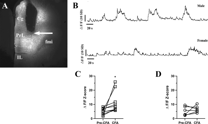
Figure 2: Inflammatory pain increases the neuronal activity of the mPFC neurons in freely moving male mice but not in female mice.
Panel A shows the placement of the photometric fiber-optic cannula in the mPFC. The arrow points to the cannula end positioned above the transduced neurons occupying the deep layers of the PrL, a subdivision of mPFC. Panel B shows the continuous fiber photometry recordings from a male (upper trace) and female (lower trace) mice. The mice ambulated on elevated O-maze during the recordings, eight days after CFA injection into the hind paw. The graph in panel C is the mean z-scored change in fluorescent signal (ΛF/F) of male mice after eight days with inflammatory pain, where the CFA injection caused significant increase of the detected fluorescence triggered by Ca2+ transients (Paired T-test, t8 = 2.4, P < 0.05). Panel D is the mean z-scored change in ΛF/F of female mice with inflammatory pain. The CFA injection did not change the intensity of the fluorescent signal (Paired T-test, t5 = 0.9, P > 0.05). Data expressed as individual points. Abbreviations: Cg – cingulate cortex, fmi – forceps minor of the corpus callosum, IL – infralimbic cortex, PrL – prelimbic cortex.

